2R40

Crystal structure of 20E bound EcR/USP


Experimental Data Snapshot

  • Method: X-RAY DIFFRACTION
  • Resolution: 2.40 Å
  • R-Value Free: 0.282 
  • R-Value Work: 0.224 
  • R-Value Observed: 0.230 

wwPDB Validation   3D Report Full Report


Ligand Structure Quality Assessment 


This is version 1.3 of the entry. See complete history


Literature

Critical Role of Desolvation in the Binding of 20-Hydroxyecdysone to the Ecdysone Receptor

Browning, C.Martin, E.Loch, C.Wurtz, J.M.Moras, D.Stote, R.H.Dejaegere, A.P.Billas, I.M.L.

(2007) J Biol Chem 282: 32924-32934

  • DOI: https://doi.org/10.1074/jbc.M705559200
  • Primary Citation of Related Structures:  
    2R40

  • PubMed Abstract: 

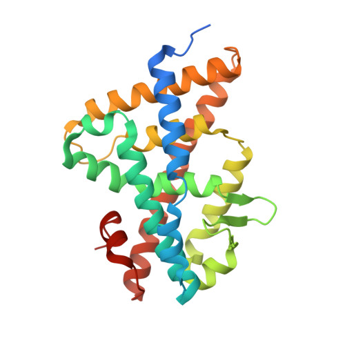
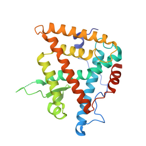
    The insect steroid hormone 20-hydroxyecdysone (20E) binds to its cognate nuclear receptor composed of the ecdysone receptor (EcR) and Ultraspiracle (USP) and triggers the main developmental transitions, in particular molting and metamorphosis. We present the crystal structure of the ligand-binding domains of EcR/USP in complex with 20E at 2.4A resolution and compare it with published structures of EcR/USP bound to ponasterone A (ponA). ponA is essentially identical to 20E but lacks the 25-OH group of 20E. The structure of 20E-bound EcR indicates that an additional hydrogen bond is formed compared with the ponA-bound receptor, yet, paradoxically, ponA has a significantly higher affinity for EcR than 20E. Theoretical studies based on docking and free energy methods lead to a rationale for understanding the difference in binding affinities between 20E and ponA. Results of the calculations indicate that the favorable contribution from the extra H-bond made by 25-OH of 20E is counterbalanced by its larger desolvation cost compared with that of ponA. The contribution of 25-OH to the binding affinity is further compared with those of 20- and 22-OH groups. Ligands that lack the 20- or 22-OH group are indeed known to bind less favorably to EcR than 20E, an effect opposite to that observed for ponA. The results indicate that their respective contributions to receptor-ligand complex stability reside mostly in their different contributions to solvation/desolvation. Together, the data demonstrate the critical role of ligand desolvation in determining binding affinity, with general implications for the binding of hormones to their cognate nuclear receptors.


  • Organizational Affiliation

    Département de Biologie et de Génomique Structurales, Institut de Génétique et de Biologie Moléculaire et Cellulaire, Ecole Supérieure de Biotechnologie de Strasbourg, Boulevard Sébastien Brant, Illkirch, France.


Macromolecules
Find similar proteins by:  (by identity cutoff)  |  3D Structure
Entity ID: 1
MoleculeChains Sequence LengthOrganismDetailsImage
Ecdysone ReceptorA [auth D]266Heliothis virescensMutation(s): 0 
Gene Names: EcR
UniProt
Find proteins for O18473 (Heliothis virescens)
Explore O18473 
Go to UniProtKB:  O18473
Entity Groups  
Sequence Clusters30% Identity50% Identity70% Identity90% Identity95% Identity100% Identity
UniProt GroupO18473
Sequence Annotations
Expand
  • Reference Sequence
Find similar proteins by:  (by identity cutoff)  |  3D Structure
Entity ID: 2
MoleculeChains Sequence LengthOrganismDetailsImage
UltraspiracleB [auth A]263Heliothis virescensMutation(s): 0 
UniProt
Find proteins for Q7SIF6 (Heliothis virescens)
Explore Q7SIF6 
Go to UniProtKB:  Q7SIF6
Entity Groups  
Sequence Clusters30% Identity50% Identity70% Identity90% Identity95% Identity100% Identity
UniProt GroupQ7SIF6
Sequence Annotations
Expand
  • Reference Sequence
Small Molecules
Ligands 5 Unique
IDChains Name / Formula / InChI Key2D Diagram3D Interactions
EPH
Query on EPH

Download Ideal Coordinates CCD File 
G [auth A]L-ALPHA-PHOSPHATIDYL-BETA-OLEOYL-GAMMA-PALMITOYL-PHOSPHATIDYLETHANOLAMINE
C39 H68 N O8 P
MABRTXOVHMDVAT-AAEGOEIASA-N
20E
Query on 20E

Download Ideal Coordinates CCD File 
E [auth D](2beta,3beta,5beta,22R)-2,3,14,20,22,25-hexahydroxycholest-7-en-6-one
C27 H44 O7
NKDFYOWSKOHCCO-YPVLXUMRSA-N
FLC
Query on FLC

Download Ideal Coordinates CCD File 
C [auth D]CITRATE ANION
C6 H5 O7
KRKNYBCHXYNGOX-UHFFFAOYSA-K
SO4
Query on SO4

Download Ideal Coordinates CCD File 
D
SULFATE ION
O4 S
QAOWNCQODCNURD-UHFFFAOYSA-L
GOL
Query on GOL

Download Ideal Coordinates CCD File 
F [auth D]GLYCEROL
C3 H8 O3
PEDCQBHIVMGVHV-UHFFFAOYSA-N
Experimental Data & Validation

Experimental Data

  • Method: X-RAY DIFFRACTION
  • Resolution: 2.40 Å
  • R-Value Free: 0.282 
  • R-Value Work: 0.224 
  • R-Value Observed: 0.230 
  • Space Group: P 31 2 1
Unit Cell:
Length ( Å )Angle ( ˚ )
a = 58.001α = 90
b = 58.001β = 90
c = 303.592γ = 120
Software Package:
Software NamePurpose
DENZOdata reduction
SCALEPACKdata scaling
REFMACrefinement
PDB_EXTRACTdata extraction
ADSCdata collection
AMoREphasing

Structure Validation

View Full Validation Report


Ligand Structure Quality Assessment 


Entry History 

Deposition Data

Revision History  (Full details and data files)

  • Version 1.0: 2007-10-02
    Type: Initial release
  • Version 1.1: 2011-07-13
    Changes: Advisory, Refinement description, Version format compliance
  • Version 1.2: 2017-10-25
    Changes: Refinement description
  • Version 1.3: 2024-02-21
    Changes: Data collection, Database references, Derived calculations