Structural basis for the geometry-driven localization of a small protein.
Gill, R.L., Castaing, J.P., Hsin, J., Tan, I.S., Wang, X., Huang, K.C., Tian, F., Ramamurthi, K.S.(2015) Proc Natl Acad Sci U S A 112: E1908-E1915
- PubMed: 25825747
- DOI: https://doi.org/10.1073/pnas.1423868112
- Primary Citation of Related Structures:
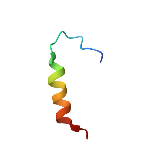
2MVH, 2MVJ - PubMed Abstract:
In bacteria, certain shape-sensing proteins localize to differently curved membranes. During sporulation in Bacillus subtilis, the only convex (positively curved) surface in the cell is the forespore, an approximately spherical internal organelle. Previously, we demonstrated that SpoVM localizes to the forespore by preferentially adsorbing onto slightly convex membranes. Here, we used NMR and molecular dynamics simulations of SpoVM and a localization mutant (SpoVM(P9A)) to reveal that SpoVM's atypical amphipathic α-helix inserts deeply into the membrane and interacts extensively with acyl chains to sense packing differences in differently curved membranes. Based on binding to spherical supported lipid bilayers and Monte Carlo simulations, we hypothesize that SpoVM's membrane insertion, along with potential cooperative interactions with other SpoVM molecules in the lipid bilayer, drives its preferential localization onto slightly convex membranes. Such a mechanism, which is distinct from that used by high curvature-sensing proteins, may be widely conserved for the localization of proteins onto the surface of cellular organelles.
- Department of Biochemistry and Molecular Biology, College of Medicine, Pennsylvania State University, Hershey, PA 17033;
Organizational Affiliation: