Negatively charged lipid membranes promote a disorder-order transition in the Yersinia YscU protein.
Weise, C.F., Login, F.H., Ho, O., Grobner, G., Wolf-Watz, H., Wolf-Watz, M.(2014) Biophys J 107: 1950-1961
- PubMed: 25418176
- DOI: https://doi.org/10.1016/j.bpj.2014.09.005
- Primary Citation of Related Structures:
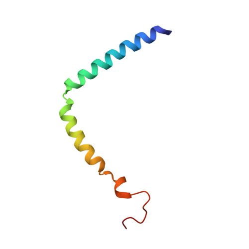
2ML9 - PubMed Abstract:
The inner membrane of Gram-negative bacteria is negatively charged, rendering positively charged cytoplasmic proteins in close proximity likely candidates for protein-membrane interactions. YscU is a Yersinia pseudotuberculosis type III secretion system protein crucial for bacterial pathogenesis. The protein contains a highly conserved positively charged linker sequence that separates membrane-spanning and cytoplasmic (YscUC) domains. Although disordered in solution, inspection of the primary sequence of the linker reveals that positively charged residues are separated with a typical helical periodicity. Here, we demonstrate that the linker sequence of YscU undergoes a largely electrostatically driven coil-to-helix transition upon binding to negatively charged membrane interfaces. Using membrane-mimicking sodium dodecyl sulfate micelles, an NMR derived structural model reveals the induction of three helical segments in the linker. The overall linker placement in sodium dodecyl sulfate micelles was identified by NMR experiments including paramagnetic relaxation enhancements. Partitioning of individual residues agrees with their hydrophobicity and supports an interfacial positioning of the helices. Replacement of positively charged linker residues with alanine resulted in YscUC variants displaying attenuated membrane-binding affinities, suggesting that the membrane interaction depends on positive charges within the linker. In vivo experiments with bacteria expressing these YscU replacements resulted in phenotypes displaying significantly reduced effector protein secretion levels. Taken together, our data identify a previously unknown membrane-interacting surface of YscUC that, when perturbed by mutations, disrupts the function of the pathogenic machinery in Yersinia.
- Department of Chemistry, Chemical Biological Center, Umeå University, Umeå, Sweden.
Organizational Affiliation: