Solution structure of recombinant somatomedin B domain from vitronectin produced in Pichia pastoris
Kjaergaard, M., Gardsvoll, H., Hirschberg, D., Nielbo, S., Mayasundari, A., Peterson, C.B., Jansson, A., Jorgensen, T.J.D., Poulsen, F.M., Ploug, M.(2007) Protein Sci 16: 1934-1945
- PubMed: 17766387
- DOI: https://doi.org/10.1110/ps.072949607
- Primary Citation of Related Structures:
2JQ8 - PubMed Abstract:
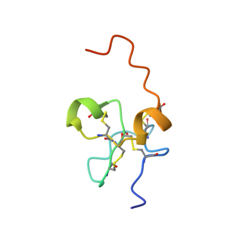
The cysteine-rich somatomedin B domain (SMB) of the matrix protein vitronectin is involved in several important biological processes. First, it stabilizes the active conformation of the plasminogen activator inhibitor (PAI-1); second, it provides the recognition motif for cell adhesion via the cognate integrins (alpha(v)beta(3), alpha(v)beta(5), and alpha(IIb)beta(3)); and third, it binds the complex between urokinase-type plasminogen activator (uPA) and its glycolipid-anchored receptor (uPAR). Previous structural studies on SMB have used recombinant protein expressed in Escherichia coli or SMB released from plasma-derived vitronectin by CNBr cleavage. However, different disulfide patterns and three-dimensional structures for SMB were reported. In the present study, we have expressed recombinant human SMB by two different eukaryotic expression systems, Pichia pastoris and Drosophila melanogaster S2-cells, both yielding structurally and functionally homogeneous protein preparations. Importantly, the entire population of our purified, recombinant SMB has a solvent exposure, both as a free domain and in complex with PAI-1, which is indistinguishable from that of plasma-derived SMB as assessed by amide hydrogen ((1)H/(2)H) exchange. This solvent exposure was only reproduced by one of three synthetic SMB products with predefined disulfide connectivities corresponding to those published previously. Furthermore, this connectivity was also the only one to yield a folded and functional domain. The NMR structure was determined for free SMB produced by Pichia and is largely consistent with that solved by X-ray crystallography for SMB in complex with PAI-1.
- Finsen Laboratory, Rigshospitalet Section 3735, Copenhagen Biocenter, DK-2200 Copenhagen N, Denmark.
Organizational Affiliation: