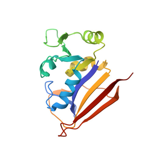
NMR structures of apo L.casei dihydrofolate reductase and its complexes with trimethoprim and NADPH. Contributions to positive cooperative binding from ligand-induced refolding, conformational changes and interligand hydrophobic interactions
Feeney, J., Birdsall, B., Kovalevskaya, N.V., Smurnyy, Y.D., Navarro-Peran, E.M., Polshakov, V.I.(2011) Biochemistry
- PubMed: 21410224
- DOI: https://doi.org/10.1021/bi200067t
- Primary Citation of Related Structures:
2HM9, 2L28, 2LF1 - PubMed Abstract:
In order to examine the origins of the large positive cooperativity (ΔG(0)(coop) = -2.9 kcal mol(-1)) of trimethoprim (TMP) binding to a bacterial dihydrofolate reductase (DHFR) in the presence of NADPH, we have determined and compared NMR solution structures of L. casei apo DHFR and its binary and ternary complexes with TMP and NADPH and made complementary thermodynamic measurements. The DHFR structures are generally very similar except for the A-B loop region and part of helix B (residues 15-31) which could not be directly detected for L. casei apo DHFR because of line broadening from exchange between folded and unfolded forms. Thermodynamic and NMR measurements suggested that a significant contribution to the cooperativity comes from refolding of apo DHFR on binding the first ligand (up to -0.95 kcals mol(-1) if 80% of A-B loop requires refolding). Comparisons of Cα-Cα distance differences and domain rotation angles between apo DHFR and its complexes indicated that generally similar conformational changes involving domain movements accompany formation of the binary complexes with either TMP or NADPH and that the binary structures are approaching that of the ternary complex as would be expected for positive cooperativity. These favorable ligand-induced structural changes upon binding the first ligand will also contribute significantly to the cooperative binding. A further substantial contribution to cooperative binding results from the proximity of the bound ligands in the ternary complex: this reduces the solvent accessible area of the ligand and provides a favorable entropic hydrophobic contribution (up to -1.4 kcal mol(-1)).
- Division of Molecular Structure, MRC National Institute for Medical Research, The Ridgeway, Mill Hill, London NW7 1AA, U.K. jfeeney@nimr.mrc.ac.uk
Organizational Affiliation: