2AK7

structure of a dimeric P-Ser-Crh


Experimental Data Snapshot

  • Method: X-RAY DIFFRACTION
  • Resolution: 2.00 Å
  • R-Value Free: 0.214 
  • R-Value Work: 0.199 

wwPDB Validation   3D Report Full Report


This is version 1.3 of the entry. See complete history


Literature

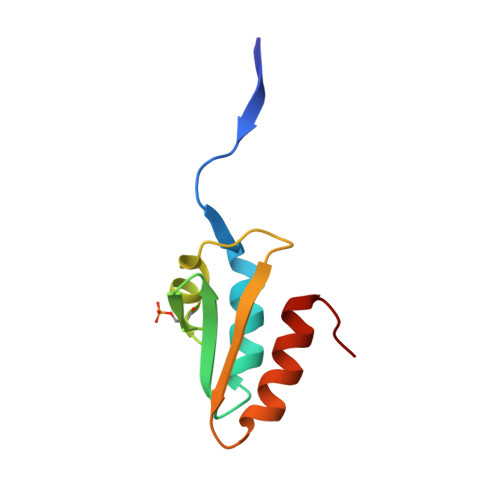
X-ray structure of a domain-swapped dimer of Ser46-phosphorylated Crh from Bacillus subtilis.

Chaptal, V.Lariviere, L.Gueguen-Chaignon, V.Galinier, A.Nessler, S.Morera, S.

(2006) Proteins 63: 249-251


Macromolecules
Find similar proteins by:  (by identity cutoff)  |  3D Structure
Entity ID: 1
MoleculeChains Sequence LengthOrganismDetailsImage
HPr-like protein crh
A, B
86Bacillus subtilisMutation(s): 1 
Gene Names: crh
UniProt
Find proteins for O06976 (Bacillus subtilis (strain 168))
Explore O06976 
Go to UniProtKB:  O06976
Entity Groups  
Sequence Clusters30% Identity50% Identity70% Identity90% Identity95% Identity100% Identity
UniProt GroupO06976
Sequence Annotations
Expand
  • Reference Sequence
Small Molecules
Modified Residues  1 Unique
IDChains TypeFormula2D DiagramParent
SEP
Query on SEP
A, B
L-PEPTIDE LINKINGC3 H8 N O6 PSER
Experimental Data & Validation

Experimental Data

  • Method: X-RAY DIFFRACTION
  • Resolution: 2.00 Å
  • R-Value Free: 0.214 
  • R-Value Work: 0.199 
  • Space Group: P 43 2 2
Unit Cell:
Length ( Å )Angle ( ˚ )
a = 67.697α = 90
b = 67.697β = 90
c = 117.157γ = 90
Software Package:
Software NamePurpose
DENZOdata reduction
SCALEPACKdata scaling
AMoREphasing
CNSrefinement

Structure Validation

View Full Validation Report


Entry History 

Deposition Data

Revision History  (Full details and data files)

  • Version 1.0: 2006-06-27
    Type: Initial release
  • Version 1.1: 2008-04-30
    Changes: Version format compliance
  • Version 1.2: 2011-07-13
    Changes: Version format compliance
  • Version 1.3: 2023-08-23
    Changes: Data collection, Database references, Derived calculations, Refinement description