29MV | pdb_000029mv

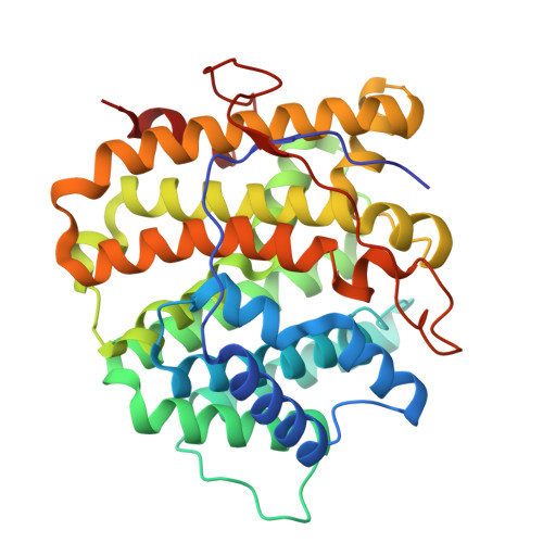
Sesterterpene Synthase from Streptomyces subrutilus (Subrutilane Synthase, SrS) in complex with the surrogate geranyl-farnesyl-thiopyrophosphate (GFSPP)


Experimental Data Snapshot

  • Method: X-RAY DIFFRACTION
  • Resolution: 2.30 Å
  • R-Value Free: 
    0.211 (Depositor), 0.216 (DCC) 
  • R-Value Work: 
    0.179 (Depositor), 0.189 (DCC) 
  • R-Value Observed: 
    0.181 (Depositor) 

Starting Model: in silico
View more details

wwPDB Validation 3D Report Full Report

Validation slider image for 29MV

Ligand Structure Quality Assessment 


This is version 1.1 of the entry. See complete history

Literature

Local Active-Site Architecture Directs Divergent Carbocation Cascades in Sesterterpene Synthases.

Troycke, P.Li, H.Yang, K.Dickschat, J.S.Groll, M.

(2026) J Am Chem Soc 148: 36648-36661

  • DOI: https://doi.org/10.1021/jacs.6c06677
  • Primary Citation Related Structures: 
    29MK, 29MQ, 29MS, 29MT, 29MU, 29MV

  • PubMed Abstract: 

    Type I terpene synthases generate complex polycyclic scaffolds through carbocation cascades. However, how closely related enzymes convert a common C25 precursor to distinct sesterterpene frameworks remains unresolved. Here, we combine high-resolution crystal structures with systematic mutagenesis of four bacterial sesterterpene synthases to define the structural basis for pathway divergence. These analyses show how local active-site interactions within a conserved fold redirect carbocation trajectories and thereby control product formation. The structures reveal a preorganized binding mode that positions the substrate in a product-like conformation and directs the cyclization cascade. Comparative structural analyses further support distinct carbocation trajectories involving either centralized cation hubs or sequential rearrangement pathways with the exact pathway being enzyme-dependent. Structure-guided mutagenesis targets these features, alters product profiles, and enables the formation of new terpene scaffolds. Together, crystallographic data and mutagenesis demonstrate that closely related enzymes with the same overall fold can follow distinct carbocation trajectories and reveal how local architectural changes control the cyclization outcome.


  • Organizational Affiliation
    • Center for Protein Assemblies, Department of Bioscience, School of Natural Sciences, Technical University Munich, 85748Garching, Germany.

Macromolecule Content 

  • Total Structure Weight: 162.27 kDa 
  • Atom Count: 11,081 
  • Modeled Residue Count: 1,350 
  • Deposited Residue Count: 1,428 
  • Unique protein chains: 1

Macromolecules

Find similar proteins by:|  3D Structure
Entity ID: 1
MoleculeChains  Sequence LengthOrganismDetailsImage
Terpene synthase
A, B, C, D
357Streptomyces subrutilusMutation(s): 0 
Gene Names: CP968_31920GCM10010371_68870
EC: 4.2.3
UniProt
Find proteins for A0A5P2UZR8 (Streptomyces subrutilus)
Explore A0A5P2UZR8 
Go to UniProtKB:  A0A5P2UZR8
Entity Groups
Sequence Clusters30% Identity50% Identity70% Identity90% Identity95% Identity100% Identity
UniProt GroupA0A5P2UZR8
Sequence Annotations
Expand
Reference Sequence

Small Molecules

Ligands 4 Unique
IDChains Name / Formula / InChI Key2D Diagram3D Interactions
A1JI6

Query on A1JI6


Download:Ideal Coordinates CCD File
E [auth A][(2~{E},6~{E},10~{E},14~{E})-3,7,11,15,19-pentamethylicosa-2,6,10,14,18-pentaenyl]sulfanyl-phosphonooxy-phosphinic acid
C25 H44 O6 P2 S
GLTFGFJVZBHKFR-GIXZANJISA-N
PEG
(Subject of Investigation/LOI)

Query on PEG


Download:Ideal Coordinates CCD File
G [auth A]
K [auth C]
L [auth C]
M [auth C]
O [auth D]
G [auth A],
K [auth C],
L [auth C],
M [auth C],
O [auth D],
P [auth D]
DI(HYDROXYETHYL)ETHER
C4 H10 O3
MTHSVFCYNBDYFN-UHFFFAOYSA-N
GOL
(Subject of Investigation/LOI)

Query on GOL


Download:Ideal Coordinates CCD File
F [auth A],
N [auth D]
GLYCEROL
C3 H8 O3
PEDCQBHIVMGVHV-UHFFFAOYSA-N
MG

Query on MG


Download:Ideal Coordinates CCD File
H [auth A],
I [auth A],
J [auth A]
MAGNESIUM ION
Mg
JLVVSXFLKOJNIY-UHFFFAOYSA-N

Experimental Data & Validation

Experimental Data

  • Method: X-RAY DIFFRACTION
  • Resolution: 2.30 Å
  • R-Value Free:  0.211 (Depositor), 0.216 (DCC) 
  • R-Value Work:  0.179 (Depositor), 0.189 (DCC) 
  • R-Value Observed: 0.181 (Depositor) 
Space Group: P 1 21 1
Unit Cell:
Length ( Å )Angle ( ˚ )
a = 74.91α = 90
b = 89.63β = 91.1
c = 102.69γ = 90
Software Package:
Software NamePurpose
REFMACrefinement
PDB_EXTRACTdata extraction
XDSdata reduction
XSCALEdata scaling
PHASERphasing

Structure Validation

View Full Validation Report


Ligand Structure Quality Assessment 


Entry History 

& Funding Information

Deposition Data


Funding OrganizationLocationGrant Number
German Research Foundation (DFG)GermanyGR 1861/13-1 (project number 542938137)

Revision History  (Full details and data files)

  • Version 1.0: 2026-09-02
    Type: Initial release
  • Version 1.1: 2026-09-16
    Changes: Database references