29LJ | pdb_000029lj

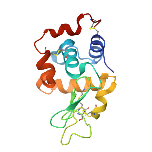
X-ray structure of the adduct formed upon reaction of Lysozyme with [Ru2Cl(D-p-CNPhF)(O2CCH3)3]


Experimental Data Snapshot

  • Method: X-RAY DIFFRACTION
  • Resolution: 1.13 Å
  • R-Value Free: 
    0.209 (Depositor), 0.215 (DCC) 
  • R-Value Work: 
    0.198 (Depositor), 0.205 (DCC) 

Starting Model: experimental
View more details

wwPDB Validation 3D Report Full Report

Validation slider image for 29LJ

Ligand Structure Quality Assessment 


This is version 1.1 of the entry. See complete history

Literature

Protein Recognition and Amyloid Remodeling Governed by Paddlewheel Diruthenium Coordination Chemistry.

La Manna, S.Florio, D.Ferraro, G.Policino, M.R.Altieri, F.Teran, A.Herrero, S.Merlino, A.Marasco, D.

(2026) Inorg Chem 65: 19953-19964

  • DOI: https://doi.org/10.1021/acs.inorgchem.6c02947
  • Primary Citation Related Structures: 
    29LJ, 29MN, 29MR, 29MY, 29NX

  • PubMed Abstract: 

    Paddlewheel diruthenium (Ru2) complexes are promising modulators of protein aggregation due to their tunable coordination chemistry and dual-action properties. Here, we investigate the interaction of five Ru2 complexes with hen egg white lysozyme (HEWL), an amyloid model, to elucidate their antiaggregation capabilities. High-resolution X-ray crystallography shows that all complexes preferentially bind to Asp119 and, in some cases, Asp101, through coordination to the Ru2 core while preserving the overall protein fold. Both covalent and noncovalent interactions are observed, depending on ligand environment and steric effects. Solution studies confirm the formation of HEWL-Ru2 adducts under both neutral and acidic conditions. Functional assays demonstrate that all Ru2 complexes effectively inhibit HEWL fibrillogenesis, as indicated by reduced ThT fluorescence and the absence of large aggregates in dynamic light scattering measurements. Disaggregation of preformed fibrils was more variable, with the complex bearing vacant axial sites showing the highest activity. Circular dichroism and scanning electron microscopy analyses reveal that these compounds redirect aggregation toward noncanonical morphologies rather than fully dissolving fibrils. Cytotoxicity assays confirm reduced HEWL-induced cellular toxicity. Overall, our findings establish a correlation between ligand composition, coordination behavior, protein binding, and antiamyloid activity, providing a framework for designing Ru2-based multifunctional modulators of protein aggregation.


  • Organizational Affiliation
    • Department of Pharmacy, University of Naples Federico II, 80131Naples, Italy.

Macromolecule Content 

  • Total Structure Weight: 15.86 kDa 
  • Atom Count: 1,266 
  • Modeled Residue Count: 129 
  • Deposited Residue Count: 129 
  • Unique protein chains: 1

Macromolecules

Find similar proteins by:|  3D Structure
Entity ID: 1
MoleculeChains  Sequence LengthOrganismDetailsImage
Lysozyme C129Gallus gallusMutation(s): 0 
EC: 3.2.1.17
UniProt
Find proteins for P00698 (Gallus gallus)
Explore P00698 
Go to UniProtKB:  P00698
Entity Groups
Sequence Clusters30% Identity50% Identity70% Identity90% Identity95% Identity100% Identity
UniProt GroupP00698
Sequence Annotations
Expand
Reference Sequence

Small Molecules

Ligands 4 Unique
IDChains Name / Formula / InChI Key2D Diagram3D Interactions
A1IHW
(Subject of Investigation/LOI)

Query on A1IHW


Download:Ideal Coordinates CCD File
H [auth A],
I [auth A]
4-[11-(4-cyanophenyl)-1,5-bis($l^{3}-oxidanyl)-3,7-dimethyl-2$l^{3},4,6,8$l^{3}-tetraoxa-9$l^{4},11-diaza-1$l^{5},5$l^{5}-diruthenatricyclo[3.3.3.0^{1,5}]undeca-2,7,9-trien-9-yl]benzenecarbonitrile
C19 H19 N4 O6 Ru2
LPPYWRVZKKMORQ-UHFFFAOYSA-L
NO3

Query on NO3


Download:Ideal Coordinates CCD File
C [auth A],
D [auth A],
E [auth A],
F [auth A]
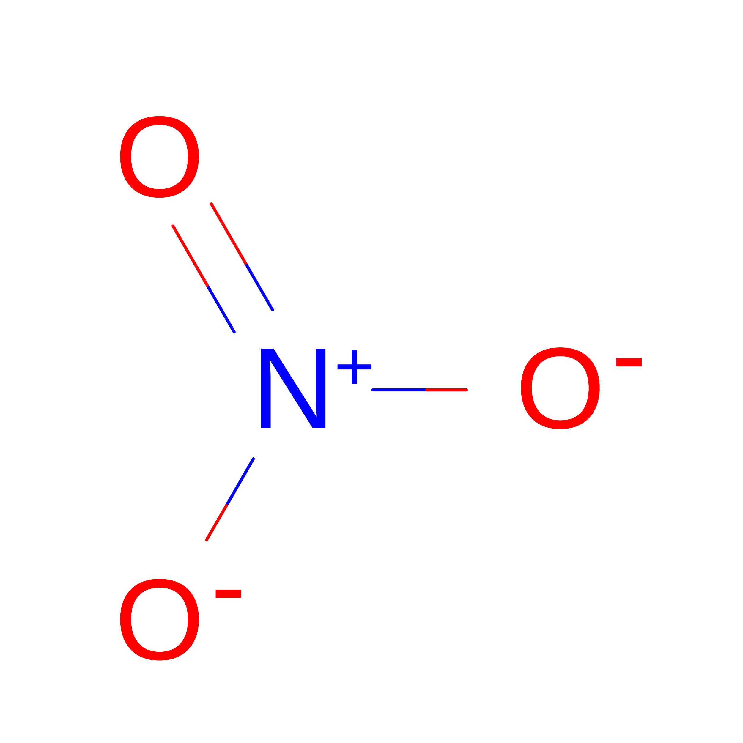
NITRATE ION
N O3
NHNBFGGVMKEFGY-UHFFFAOYSA-N
ACT

Query on ACT


Download:Ideal Coordinates CCD File
G [auth A]ACETATE ION
C2 H3 O2
QTBSBXVTEAMEQO-UHFFFAOYSA-M
NA

Query on NA


Download:Ideal Coordinates CCD File
B [auth A]SODIUM ION
Na
FKNQFGJONOIPTF-UHFFFAOYSA-N

Experimental Data & Validation

Experimental Data

  • Method: X-RAY DIFFRACTION
  • Resolution: 1.13 Å
  • R-Value Free:  0.209 (Depositor), 0.215 (DCC) 
  • R-Value Work:  0.198 (Depositor), 0.205 (DCC) 
Space Group: P 43 21 2
Unit Cell:
Length ( Å )Angle ( ˚ )
a = 78.18α = 90
b = 78.18β = 90
c = 37.53γ = 90
Software Package:
Software NamePurpose
REFMACrefinement
autoPROCdata reduction
autoPROCdata scaling
PHASERphasing

Structure Validation

View Full Validation Report


Ligand Structure Quality Assessment 


Entry History 

& Funding Information

Deposition Data


Funding OrganizationLocationGrant Number
Not funded--

Revision History  (Full details and data files)

  • Version 1.0: 2026-09-02
    Type: Initial release
  • Version 1.1: 2026-09-16
    Changes: Database references