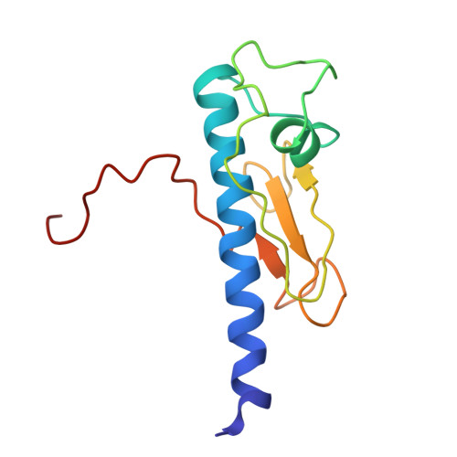
29JD | pdb_000029jd

NMR Structure of the Calcium bound form of OutG, major pilin from Dickeya dadantii T2SS


Experimental Data Snapshot

  • Method: SOLUTION NMR
  • Conformers Calculated: 50 
  • Conformers Submitted: 10 
  • Selection Criteria: structures with the lowest energy 

wwPDB Validation 3D Report Full Report

Validation slider image for 29JD

This is version 1.0 of the entry. See complete history

Literature

Structural determinants of endopilus assembly, stability, and functional specificity in bacterial type II secretion.

Lejeune, M.Ivashchenko, S.Dazzoni, R.Bardiaux, B.Sonani, R.R.Vos, M.Jacobsen, T.Egelman, E.H.Nilges, M.Francetic, O.Shevchik, V.E.Izadi-Pruneyre, N.

(2026) Structure 

  • DOI: https://doi.org/10.1016/j.str.2026.07.013
  • Primary Citation Related Structures: 
    29JD

  • PubMed Abstract: 

    Gram-negative bacteria employ the type II secretion system (T2SS) to transport folded protein effectors via a periplasmic helical polymer called the endopilus, composed of one major and four minor pilin subunits. Endopili resemble type IV pili but feature a conserved calcium-binding site stabilizing their major pilins. Endopilus polymerization is coupled to substrate translocation through a dedicated outer membrane channel. We compared T2SSs from plant and human pathogens, Dickeya dadantii and Klebsiella oxytoca, respectively. Despite different ecological niches and secreted effectors, their major pilins (OutG and PulG) share >77% sequence identity. Using NMR and cryo-EM, we solved structures of calcium-bound OutG monomer, as well as OutG and PulG endopili at 3.6 Å resolution. Combining structural, mutational, and biophysical analyses with in vivo assays, we identified key determinants of secretion specificity and endopilus stability. Our findings reveal how minor sequence variations in conserved nanomachines drive functional adaptation to diverse environments.


  • Organizational Affiliation
    • Institut Pasteur, Université Paris Cité, CNRS UMR3528, Bacterial Transmembrane Systems Unit, 75015 Paris, France.

Macromolecule Content 

  • Total Structure Weight: 13.59 kDa 
  • Atom Count: 953 
  • Modeled Residue Count: 125 
  • Deposited Residue Count: 125 
  • Unique protein chains: 1

Macromolecules

Find similar proteins by:|  3D Structure
Entity ID: 1
MoleculeChains  Sequence LengthOrganismDetailsImage
Type II secretion system core protein G125Dickeya dadantiiMutation(s): 0 
Gene Names: outGDda3937_02418
UniProt
Find proteins for E0SM38 (Dickeya dadantii (strain 3937))
Explore E0SM38 
Go to UniProtKB:  E0SM38
Entity Groups
Sequence Clusters30% Identity50% Identity70% Identity90% Identity95% Identity100% Identity
UniProt GroupE0SM38
Sequence Annotations
Expand
Reference Sequence

Small Molecules

Ligands 1 Unique
IDChains Name / Formula / InChI Key2D Diagram3D Interactions
CA
(Subject of Investigation/LOI)

Query on CA


Download:Ideal Coordinates CCD File
B [auth A]CALCIUM ION
Ca
BHPQYMZQTOCNFJ-UHFFFAOYSA-N

Experimental Data & Validation

Experimental Data

  • Method: SOLUTION NMR
  • Conformers Calculated: 50 
  • Conformers Submitted: 10 
  • Selection Criteria: structures with the lowest energy 

Structure Validation

View Full Validation Report


Entry History 

& Funding Information

Deposition Data


Funding OrganizationLocationGrant Number
H2020 Marie Curie Actions of the European CommissionEuropean Union765042
Agence Nationale de la Recherche (ANR)FranceANR-19-CE11-0020-01

Revision History  (Full details and data files)

  • Version 1.0: 2026-09-09
    Type: Initial release