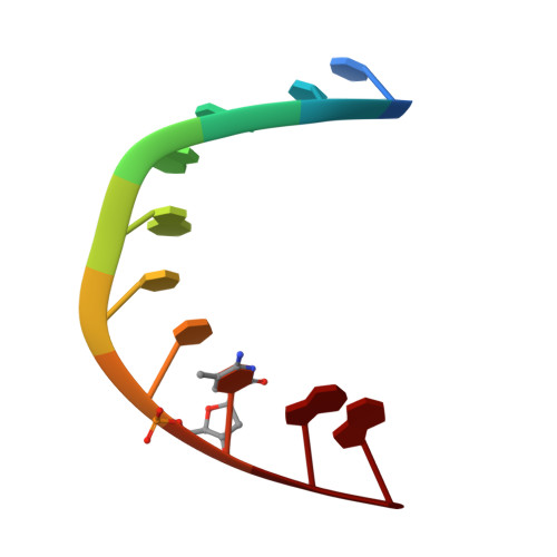
Crystal structure of the A-DNA decamer d(CCIGGCCm5CGG) at 1.6 A showing the unexpected wobble I.m5C base pair.
Ramakrishnan, B., Sundaralingam, M.(1995) Biophys J 69: 553-558
- PubMed: 8527669
- DOI: https://doi.org/10.1016/S0006-3495(95)79928-3
- Primary Citation of Related Structures:
213D - PubMed Abstract:
The crystal structure of the self-complementary decamer d(CCIGGCCm5CGG), where I and m5C replace A and T, respectively, in the Watson-Crick B-DNA decamer d(CCAGGCCTGG) is in the A-DNA conformation. Furthermore, the A-DNA duplex exhibits the unexpected wobble I.m5C+ base pairs with N3 of 5-methylcytosine protonated. The crystals belong to the orthorhombic system, space group P2(1)2(1)2(1), with a = 25.02, b = 44.95 and c = 47.62 A with one DNA duplex in the asymmetric unit. Intensity data were collected on our Siemens area detector to 1.6 A resolution. The structure was solved by the molecular placement method starting with a model from an isomorphous structure. The refinement gave a final R value of 16.3% for 404 DNA atoms and 104 water molecules using 5119 reflections. The hydration of the I.m5C+ wobble base pairs in the major groove stabilizes them as in the G.T/G.U wobbles. A comparison with the Watson-Crick base pairs of another isomorphous structure d(GCGGGCCCGC) reveals that the wobble base pairs are better described by a rotation of the individual nucleotide units around their centers of gravity. This is in contrast to the earlier description of translation of the bases into the grooves. The exposed N4 amino group of 5-methylcytosine in the wobble base pair provides a rationale for its deamination to thymine.
Organizational Affiliation:
Department of Chemistry, Ohio State University, Columbus 43210, USA.