Structure and Inter-domain Interactions of Domain II from the Blood-stage Malarial Protein, Apical Membrane Antigen 1.
Feng, Z.P., Keizer, D.W., Stevenson, R.A., Yao, S., Babon, J.J., Murphy, V.J., Anders, R.F., Norton, R.S.(2005) J Mol Biol 350: 641-656
- PubMed: 15964019
- DOI: https://doi.org/10.1016/j.jmb.2005.05.011
- Primary Citation of Related Structures:
1YXE - PubMed Abstract:
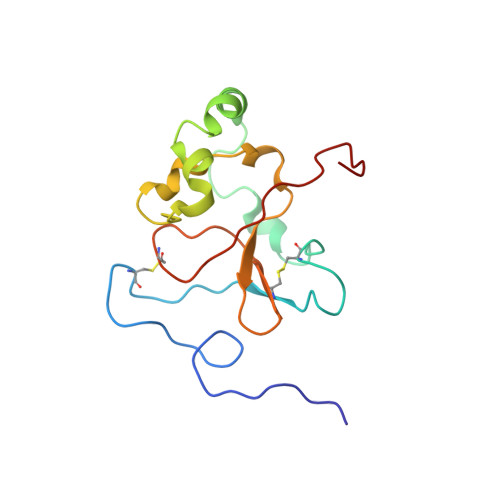
The malarial surface antigen apical membrane antigen (AMA1), from Plasmodium falciparum, is a leading candidate for inclusion in a vaccine against malaria. AMA1 is synthesised by mature blood-stages of the parasite and is located initially in the apical organelles of the merozoite. Prior to merozoite invasion of host erythrocytes, it is processed into a 66 kDa type 1 integral membrane protein on the merozoite surface. The pattern of disulphide bonds in AMA1 has been the basis for separation of the ectodomain into three domains, with three, two and three disulphide bonds, respectively. We have determined the solution structure of a 16kDa construct corresponding to the putative second domain of AMA1. While circular dichroism and hydrodynamic data were consistent with a folded structure for domain II, its NMR spectra were characterised by broad lines and significant peak overlap, more typical of a molten globule. Consistent with this, domain II bound the fluorescent dye 8-anilino-1-naphthalene sulphonate (ANS). We have nonetheless determined a structure, which defines the secondary structure elements and global fold. The two disulphide bonds link the N and C-terminal regions of the molecule, which come together to form a four-stranded beta-sheet linked to a short helix. A long loop linking the N and C-terminal regions contains four other alpha-helices, the locations of which are not fixed relative to the beta-sheet core, even though they are well-defined locally. Very recently this region of domain II has been shown to contain the epitope recognised by the invasion-inhibitory antibody 4G2, even though it does not contain any of the polymorphisms that are regarded as having arisen in response to the pressure of immune recognition.
Organizational Affiliation:
The Walter and Eliza Hall Institute of Medical Research, 1G Royal Parade, Parkville, Vic., 3050, Australia.