1YMC

THREE-DIMENSIONAL STRUCTURE OF CYANOMET-SULFMYOGLOBIN C


Experimental Data Snapshot

  • Method: X-RAY DIFFRACTION
  • Resolution: 2.00 Å
  • R-Value Observed: 0.129 

wwPDB Validation   3D Report Full Report


Ligand Structure Quality Assessment 


This is version 1.4 of the entry. See complete history


Literature

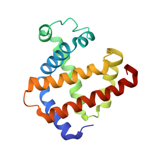
Three-dimensional structure of cyanomet-sulfmyoglobin C.

Evans, S.V.Sishta, B.P.Mauk, A.G.Brayer, G.D.

(1994) Proc Natl Acad Sci U S A 91: 4723-4726

  • DOI: https://doi.org/10.1073/pnas.91.11.4723
  • Primary Citation of Related Structures:  
    1YMC

  • PubMed Abstract: 

    The atomic structure of horse heart cyanomet-sulfmyoglobin C has been established by x-ray crystallographic techniques to a resolution of 2.0 A with an R value of 0.129. The protoheme IX prosthetic group of this thermodynamically stable sulfmyoglobin derivative has been converted to a chlorin in which the pyrrole ring bearing the 4-vinyl group is saturated and possesses an exocyclic thiolene ring. This study provides the three-dimensional structure of a protein with an iron-chlorin prosthetic group. The overall conformation of the surrounding polypeptide chain of the modified protein is very similar to that of the native protein. However, the addition of the sulfur atom has caused a distortion of the prosthetic group from that in the native protein to result in the repositioning of the side chains of some residues in the heme pocket.


  • Organizational Affiliation

    Department of Biochemistry and Molecular Biology, University of British Columbia, Vancouver, Canada.


Macromolecules
Find similar proteins by:  (by identity cutoff)  |  3D Structure
Entity ID: 1
MoleculeChains Sequence LengthOrganismDetailsImage
CYANOMET-SULFMYOGLOBIN153Equus caballusMutation(s): 0 
UniProt
Find proteins for P68082 (Equus caballus)
Explore P68082 
Go to UniProtKB:  P68082
Entity Groups  
Sequence Clusters30% Identity50% Identity70% Identity90% Identity95% Identity100% Identity
UniProt GroupP68082
Sequence Annotations
Expand
  • Reference Sequence
Experimental Data & Validation

Experimental Data

  • Method: X-RAY DIFFRACTION
  • Resolution: 2.00 Å
  • R-Value Observed: 0.129 
  • Space Group: P 1 21 1
Unit Cell:
Length ( Å )Angle ( ˚ )
a = 64.5α = 90
b = 28.91β = 107
c = 35.66γ = 90
Software Package:
Software NamePurpose
PROLSQrefinement

Structure Validation

View Full Validation Report


Ligand Structure Quality Assessment 


Entry History 

Deposition Data

Revision History  (Full details and data files)

  • Version 1.0: 1994-01-31
    Type: Initial release
  • Version 1.1: 2008-03-24
    Changes: Version format compliance
  • Version 1.2: 2011-07-13
    Changes: Version format compliance
  • Version 1.3: 2017-11-29
    Changes: Derived calculations, Other
  • Version 1.4: 2024-02-14
    Changes: Data collection, Database references, Derived calculations