1YCL

Crystal Structure of B. subtilis LuxS in Complex with a Catalytic 2-Ketone Intermediate


Experimental Data Snapshot

  • Method: X-RAY DIFFRACTION
  • Resolution: 1.80 Å
  • R-Value Free: 0.256 
  • R-Value Work: 0.225 
  • R-Value Observed: 0.225 

wwPDB Validation   3D Report Full Report


Ligand Structure Quality Assessment 


This is version 1.5 of the entry. See complete history


Literature

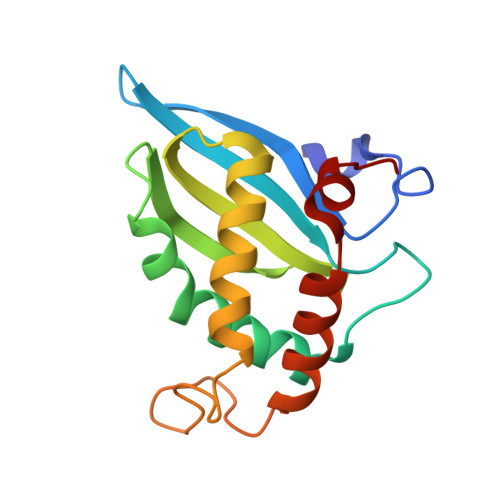
Crystal Structure of S-Ribosylhomocysteinase (LuxS) in Complex with a Catalytic 2-Ketone Intermediate.

Rajan, R.Zhu, J.Hu, X.Pei, D.Bell, C.E.

(2005) Biochemistry 44: 3745-3753

  • DOI: https://doi.org/10.1021/bi0477384
  • Primary Citation of Related Structures:  
    1YCL

  • PubMed Abstract: 

    S-Ribosylhomocysteinase (LuxS) is an Fe(2+)-dependent metalloenzyme that catalyzes the cleavage of the thioether bond in S-ribosylhomocysteine (SRH) to produce homocysteine (Hcys) and 4,5-dihydroxy-2,3-pentanedione (DPD), the precursor of type II bacterial quorum-sensing molecule. The proposed mechanism involves an initial metal-catalyzed aldose-ketose isomerization reaction, which results in the migration of the ribose carbonyl group from its C1 to C2 position and the formation of a 2-ketone intermediate. A repetition of the isomerization reaction shifts the carbonyl group to the C3 position. Subsequent beta-elimination reaction at the C4 and C5 positions completes the catalytic cycle. In this work, a catalytically inactive mutant (C84A) of Co(2+)-substituted Bacillus subtilis LuxS was cocrystallized with the 2-ketone intermediate and the structure was determined to 1.8 A resolution. The structure reveals that the C2 carbonyl oxygen is directly coordinated with the metal ion, providing strong support for the proposed Lewis acid function of the metal ion during catalysis. Cys-84 and Glu-57 are optimally positioned to act as general acids/bases during the isomerization and elimination reactions. In addition, Ser-6, His-11, and Arg-39 are involved in substrate/ intermediate binding through hydrogen bonding interactions. The above conclusions are further confirmed by site-directed mutagenesis and visible absorption spectroscopic studies.


  • Organizational Affiliation

    Department of Molecular and Cellular Biochemistry, Biophysics Program, The Ohio State University, Columbus, Ohio 43210, USA.


Macromolecules
Find similar proteins by:  (by identity cutoff)  |  3D Structure
Entity ID: 1
MoleculeChains Sequence LengthOrganismDetailsImage
S-ribosylhomocysteinase156Bacillus subtilisMutation(s): 1 
Gene Names: luxS
EC: 3.13.1
UniProt
Find proteins for O34667 (Bacillus subtilis (strain 168))
Explore O34667 
Go to UniProtKB:  O34667
Entity Groups  
Sequence Clusters30% Identity50% Identity70% Identity90% Identity95% Identity100% Identity
UniProt GroupO34667
Sequence Annotations
Expand
  • Reference Sequence
Small Molecules
Ligands 3 Unique
IDChains Name / Formula / InChI Key2D Diagram3D Interactions
KRI
Query on KRI

Download Ideal Coordinates CCD File 
D [auth A](S)-2-AMINO-4-[(2S,3R)-2,3,5-TRIHYDROXY-4-OXO-PENTYL]MERCAPTO-BUTYRIC ACID
C9 H17 N O6 S
QFXXRJSDEMCBPH-ARDNSNSESA-N
SO4
Query on SO4

Download Ideal Coordinates CCD File 
C [auth A]SULFATE ION
O4 S
QAOWNCQODCNURD-UHFFFAOYSA-L
CO
Query on CO

Download Ideal Coordinates CCD File 
B [auth A]COBALT (II) ION
Co
XLJKHNWPARRRJB-UHFFFAOYSA-N
Experimental Data & Validation

Experimental Data

  • Method: X-RAY DIFFRACTION
  • Resolution: 1.80 Å
  • R-Value Free: 0.256 
  • R-Value Work: 0.225 
  • R-Value Observed: 0.225 
  • Space Group: P 65 2 2
Unit Cell:
Length ( Å )Angle ( ˚ )
a = 62.53α = 90
b = 62.53β = 90
c = 149.65γ = 120
Software Package:
Software NamePurpose
CNSrefinement
CrystalCleardata reduction
CrystalCleardata scaling
CNSphasing

Structure Validation

View Full Validation Report


Ligand Structure Quality Assessment 


Entry History 

Deposition Data

Revision History  (Full details and data files)

  • Version 1.0: 2005-03-15
    Type: Initial release
  • Version 1.1: 2008-04-30
    Changes: Version format compliance
  • Version 1.2: 2011-07-13
    Changes: Derived calculations, Version format compliance
  • Version 1.3: 2021-10-20
    Changes: Database references, Derived calculations
  • Version 1.4: 2023-08-23
    Changes: Data collection, Refinement description
  • Version 1.5: 2024-03-13
    Changes: Source and taxonomy, Structure summary