


Crystal structure of apaG from Vibrio cholerae
Chang, C., Joachimiak, A., Li, H., Moy, S., Midwest Center for Structural Genomics (MCSG)To be published.
Experimental Data Snapshot
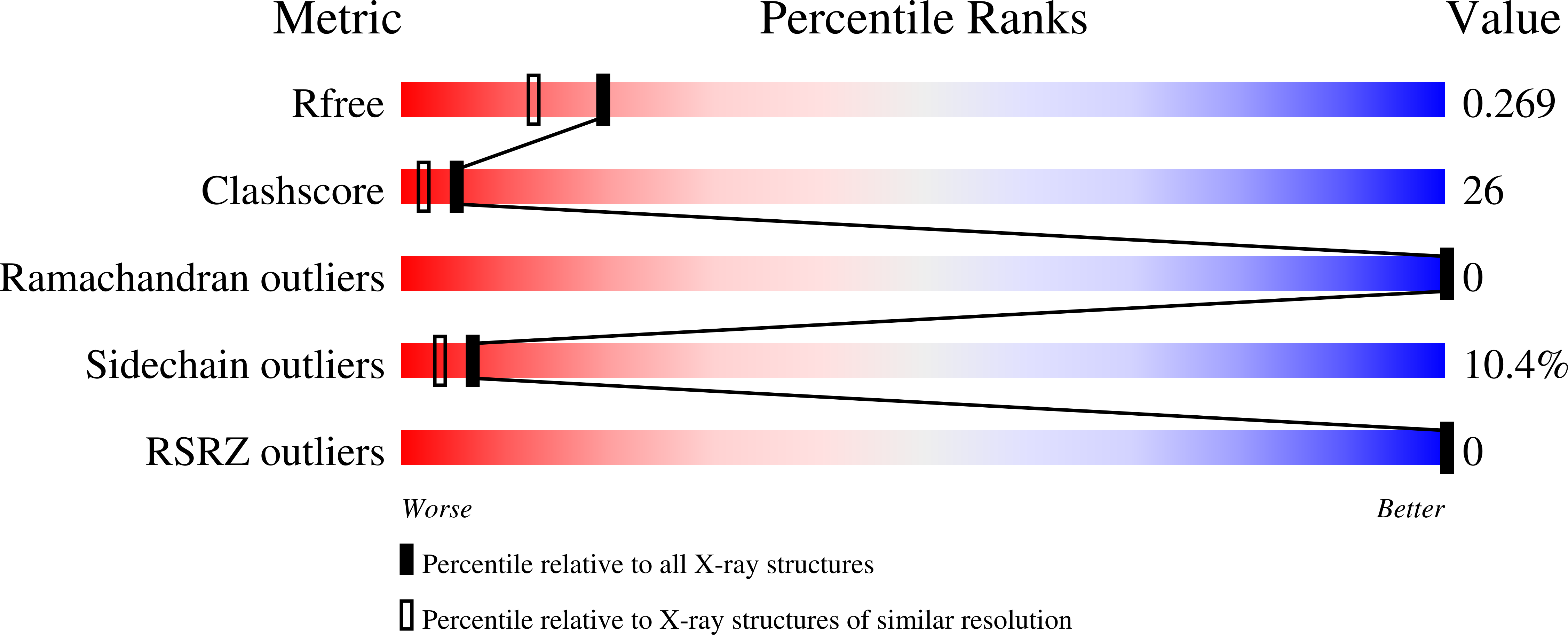
wwPDB Validation 3D Report Full Report
Entity ID: 1 | |||||
|---|---|---|---|---|---|
| Molecule | Chains | Sequence Length | Organism | Details | Image |
| Protein apaG | 126 | Vibrio cholerae | Mutation(s): 4 Gene Names: apaG | 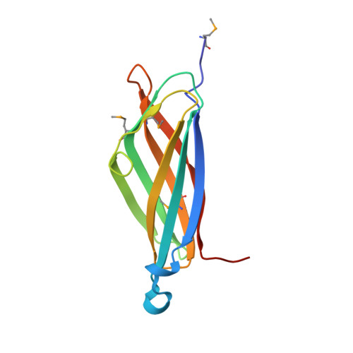 | |
UniProt | |||||
Find proteins for Q9KUS3 (Vibrio cholerae serotype O1 (strain ATCC 39315 / El Tor Inaba N16961)) Explore Q9KUS3 Go to UniProtKB: Q9KUS3 | |||||
Entity Groups | |||||
| Sequence Clusters | 30% Identity50% Identity70% Identity90% Identity95% Identity100% Identity | ||||
| UniProt Group | Q9KUS3 | ||||
Sequence AnnotationsExpand | |||||
| |||||
| Ligands 1 Unique | |||||
|---|---|---|---|---|---|
| ID | Chains | Name / Formula / InChI Key | 2D Diagram | 3D Interactions | |
| GOL Query on GOL | C [auth A] | GLYCEROL C3 H8 O3 PEDCQBHIVMGVHV-UHFFFAOYSA-N |  | ||
| Modified Residues 1 Unique | |||||
|---|---|---|---|---|---|
| ID | Chains | Type | Formula | 2D Diagram | Parent |
| MSE Query on MSE | A, B | L-PEPTIDE LINKING | C5 H11 N O2 Se |  | MET |
| Length ( Å ) | Angle ( ˚ ) |
|---|---|
| a = 90.138 | α = 90 |
| b = 67.998 | β = 131.43 |
| c = 64.002 | γ = 90 |
| Software Name | Purpose |
|---|---|
| REFMAC | refinement |
| HKL-2000 | data reduction |
| SCALEPACK | data scaling |
| AMoRE | phasing |
RCSB PDB (citation) is hosted by
RCSB PDB is a member of the

