Crystal structure of the catalytic fragment of human brain 2',3'-cyclic-nucleotide 3'-phosphodiesterase
Sakamoto, Y., Tanaka, N., Ichimiya, T., Kurihara, T., Nakamura, K.T.(2005) J Mol Biol 346: 789-800
- PubMed: 15713463
- DOI: https://doi.org/10.1016/j.jmb.2004.12.024
- Primary Citation of Related Structures:
1WOJ - PubMed Abstract:
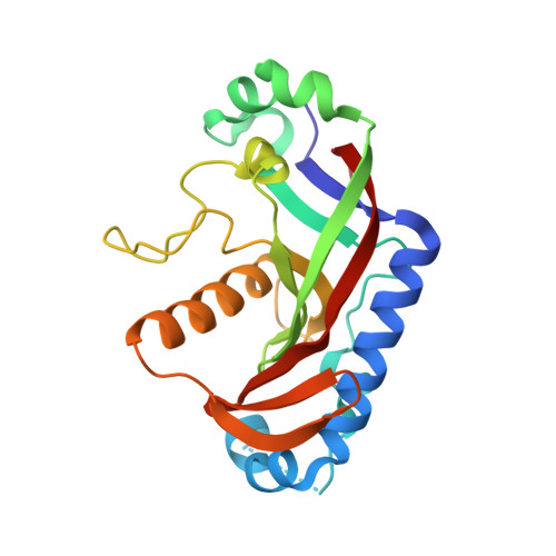
2',3'-Cyclic-nucleotide 3'-phosphodiesterase (CNP), a member of the 2H phosphoesterase superfamily, is firmly bound to brain white matter and found mainly in the central nervous system of vertebrates, and it catalyzes the hydrolysis of 2',3'-cyclic nucleotide to produce 2'-nucleotide. Recent studies on CNP-knockout mice have revealed that the absence of CNP causes axonal swelling and neuronal degeneration. Here, the crystal structure of the catalytic fragment (CF) of human CNP (hCNP-CF) is solved at 1.8A resolution. It is an alpha+beta type structure consisting of three alpha-helices and nine beta-strands. The structural core of the molecule is comprised of two topologically equivalent three-stranded antiparallel beta-sheets that are related by a pseudo 2-fold symmetry. Each beta-sheet contains an H-X-T-X motif, which is strictly conserved among members of the 2H phosphoesterase superfamily. The phosphate ion is bound to the side-chains of His and Thr from each of the two motifs. Structural comparison of hCNP-CF with plant 1'',2''-cyclic nucleotide phosphodiesterase (CPDase) and bacterial 2'-5' RNA ligase reveals that the H-X-T-X motifs are structurally conserved among these enzymes, but the surface properties of the active site are quite different among the enzymes, reflecting the differences in their substrates. On the basis of the present crystal structure of the hCNP-CF/phosphate complex, the available structure of the CPDase/cyclic-nucleotide analogue complex, and the recent functional studies of rat CNP-CF, we propose a possible substrate-binding mode and catalytic mechanism of CNP, which employs the nucleophilic water molecule activated by His310. The proposed mechanism is basically equivalent to the second step of the well-accepted reaction mechanism of RNase A. Since the overall structure of hCNP-CF differs considerably from that of RNase A, it is likely that the similar active sites with two catalytic histidine residues in these enzymes arose through convergent evolution.
Organizational Affiliation:
School of Pharmaceutical Sciences, Showa University, 1-5-8 Hatanodai, Shinagawa-ku, Tokyo 142-8555, Japan.