1W31

YEAST 5-AMINOLAEVULINIC ACID DEHYDRATASE 5-HYDROXYLAEVULINIC ACID COMPLEX


Experimental Data Snapshot

  • Method: X-RAY DIFFRACTION
  • Resolution: 1.90 Å
  • R-Value Free: 0.249 
  • R-Value Work: 0.190 

wwPDB Validation   3D Report Full Report


This is version 1.4 of the entry. See complete history


Literature

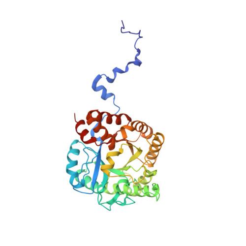
Structure of Yeast 5-Aminolaevulinic Acid Dehydratase Complexed with the Inhibitor 5-Hydroxylaevulinic Acid

Erskine, P.T.Coates, L.Newbold, R.Brindley, A.A.Stauffer, F.Beaven, G.D.E.Gill, R.Coker, A.Wood, S.P.Warren, M.J.Shoolingin-Jordan, P.M.Neier, R.Cooper, J.B.

(2005) Acta Crystallogr D Biol Crystallogr 61: 1222

  • DOI: https://doi.org/10.1107/S0907444905018834
  • Primary Citation of Related Structures:  
    1W31

  • PubMed Abstract: 

    The X-ray structure of the enzyme 5-aminolaevulinic acid dehydratase (ALAD) from yeast complexed with the competitive inhibitor 5-hydroxylaevulinic acid has been determined at a resolution of 1.9 A. The structure shows that the inhibitor is bound by a Schiff-base link to one of the invariant active-site lysine residues (Lys263). The inhibitor appears to bind in two well defined conformations and the interactions made by it suggest that it is a very close analogue of the substrate 5-aminolaevulinic acid (ALA).


  • Organizational Affiliation

    School of Biological Sciences, University of Southampton, Bassett Crescent East, Southampton SO16 7PX, England.


Macromolecules
Find similar proteins by:  (by identity cutoff)  |  3D Structure
Entity ID: 1
MoleculeChains Sequence LengthOrganismDetailsImage
DELTA-AMINOLEVULINIC ACID DEHYDRATASE342Saccharomyces cerevisiaeMutation(s): 0 
EC: 4.2.1.24
UniProt
Find proteins for P05373 (Saccharomyces cerevisiae (strain ATCC 204508 / S288c))
Explore P05373 
Go to UniProtKB:  P05373
Entity Groups  
Sequence Clusters30% Identity50% Identity70% Identity90% Identity95% Identity100% Identity
UniProt GroupP05373
Sequence Annotations
Expand
  • Reference Sequence
Small Molecules
Binding Affinity Annotations 
IDSourceBinding Affinity
SHO Binding MOAD:  1W31 Ki: 2.50e+5 (nM) from 1 assay(s)
PDBBind:  1W31 Ki: 2.50e+5 (nM) from 1 assay(s)
Experimental Data & Validation

Experimental Data

  • Method: X-RAY DIFFRACTION
  • Resolution: 1.90 Å
  • R-Value Free: 0.249 
  • R-Value Work: 0.190 
  • Space Group: I 4 2 2
Unit Cell:
Length ( Å )Angle ( ˚ )
a = 102.57α = 90
b = 102.57β = 90
c = 168.38γ = 90
Software Package:
Software NamePurpose
RESTRAINrefinement
MOSFLMdata reduction
SCALAdata scaling
CCP4phasing

Structure Validation

View Full Validation Report


Entry History 

Revision History  (Full details and data files)

  • Version 1.0: 2005-08-23
    Type: Initial release
  • Version 1.1: 2014-08-13
    Changes: Derived calculations, Other, Refinement description, Version format compliance
  • Version 1.2: 2019-05-08
    Changes: Data collection, Derived calculations, Experimental preparation, Other
  • Version 1.3: 2019-05-22
    Changes: Data collection, Refinement description
  • Version 1.4: 2023-12-13
    Changes: Data collection, Database references, Derived calculations, Other, Refinement description