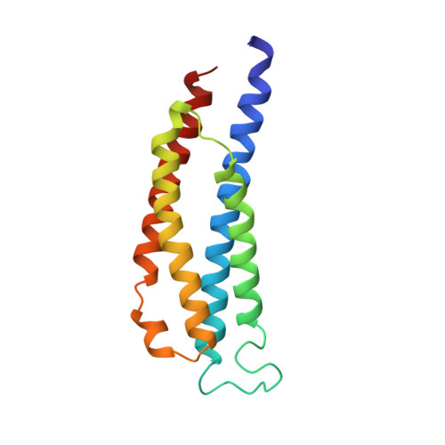
High-resolution structures of the ligand binding domain of the wild-type bacterial aspartate receptor.
Yeh, J.I., Biemann, H.P., Prive, G.G., Pandit, J., Koshland Jr., D.E., Kim, S.H.(1996) J Mol Biology 262: 186-201
- PubMed: 8831788
- DOI: https://doi.org/10.1006/jmbi.1996.0507
- Primary Citation of Related Structures:
1VLS, 1VLT - PubMed Abstract:
The high-resolution structures of the wild-type periplasmic domain of the bacterial aspartate receptor have been determined in the absence and presence of bound aspartate to 1.85 and 2.2 A resolution, respectively. As we reported earlier, in the refined structure of the complexed form of the crosslinked cysteine mutant receptor, the binding of the aspartate at the first site was mediated through four bridging water molecules while the second site showed an occupant electron density that best fit a sulfate group, which was present in the crystallization solution at high concentration. In the wild-type periplasmic domain structure two aspartate residues are bound per dimer, but with different occupancies. There exists a "strong" aspartate-binding site whose binding is again mediated by four water molecules while the second site contains aspartate whose B-factor is about 10% higher, signifying weaker binding. The interaction between the second, "weaker" aspartate with the three ligand-binding arginine side-chains is slightly different from the first site. The major difference is that there are three water molecules mediating the binding of aspartate at the second site, whereas in the first site there are four bridging water molecules. The fact that aspartate-complexed crystals of the wild-type were grown with a large excess aspartate while the cross-linked crystals were grown with equal molar aspartate may explain the difference in the stoichiometry observed. The conservation of the four bridging water molecules in the strong aspartate site of both the cross-linked and wild-type periplasmic domain may reflect an important binding motif. The periplasmic domain in the apo form is a symmetrical dimer, in which each of the subunits is equivalent, and the two aspartate binding sites are identical. Upon the binding of aspartate, the subunits are no longer symmetrical. The main difference between the aspartate-bound and unbound forms is in a small, rigid-body rotation between the subunits within a dimer. The rotation is similar in both direction and magnitude in the crosslinked and wild-type periplasmic domains. The presence of the second aspartate in the wild-type structure does not make any additional rotation compared to the single-site binding. The conservation of the small angular change in vitro suggests that the inter-subunit rotation may have relevance to the understanding of the mechanism of transmembrane signal transduction in vivo.
- Department of Chemistry, University of California Berkeley 94720, USA.
Organizational Affiliation: