1V2Z

Crystal structure of the C-terminal domain of Thermosynechococcus elongatus BP-1 KaiA


Experimental Data Snapshot

  • Method: X-RAY DIFFRACTION
  • Resolution: 1.80 Å
  • R-Value Free: 0.264 
  • R-Value Work: 0.225 
  • R-Value Observed: 0.227 

wwPDB Validation   3D Report Full Report


This is version 1.3 of the entry. See complete history


Literature

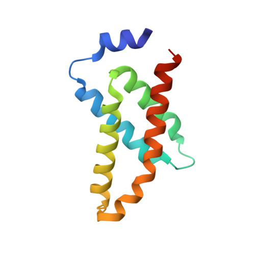
Crystal structure of the C-terminal clock-oscillator domain of the cyanobacterial KaiA protein

Uzumaki, T.Fujita, M.Nakatsu, T.Hayashi, F.Shibata, H.Itoh, N.Kato, H.Ishiura, M.

(2004) Nat Struct Mol Biol 11: 623-631

  • DOI: https://doi.org/10.1038/nsmb781
  • Primary Citation of Related Structures:  
    1V2Z

  • PubMed Abstract: 

    KaiA, KaiB and KaiC constitute the circadian clock machinery in cyanobacteria, and KaiA activates kaiBC expression whereas KaiC represses it. Here we show that KaiA is composed of three functional domains, the N-terminal amplitude-amplifier domain, the central period-adjuster domain and the C-terminal clock-oscillator domain. The C-terminal domain is responsible for dimer formation, binding to KaiC, enhancing KaiC phosphorylation and generating the circadian oscillations. The X-ray crystal structure at a resolution of 1.8 A of the C-terminal clock-oscillator domain of KaiA from the thermophilic cyanobacterium Thermosynechococcus elongatus BP-1 shows that residue His270, located at the center of a KaiA dimer concavity, is essential to KaiA function. KaiA binding to KaiC probably occurs via the concave surface. On the basis of the structure, we predict the structural roles of the residues that affect circadian oscillations.


  • Organizational Affiliation

    Center for Gene Research, Graduate School of Science, Nagoya University, Furo-cho, Chikusa-ku, Nagoya 464-8602, Japan.


Macromolecules
Find similar proteins by:  (by identity cutoff)  |  3D Structure
Entity ID: 1
MoleculeChains Sequence LengthOrganismDetailsImage
circadian clock protein KaiA homolog111Thermosynechococcus vestitus BP-1Mutation(s): 0 
UniProt
Find proteins for Q79V62 (Thermosynechococcus vestitus (strain NIES-2133 / IAM M-273 / BP-1))
Explore Q79V62 
Go to UniProtKB:  Q79V62
Entity Groups  
Sequence Clusters30% Identity50% Identity70% Identity90% Identity95% Identity100% Identity
UniProt GroupQ79V62
Sequence Annotations
Expand
  • Reference Sequence
Experimental Data & Validation

Experimental Data

  • Method: X-RAY DIFFRACTION
  • Resolution: 1.80 Å
  • R-Value Free: 0.264 
  • R-Value Work: 0.225 
  • R-Value Observed: 0.227 
  • Space Group: P 43 21 2
Unit Cell:
Length ( Å )Angle ( ˚ )
a = 61.467α = 90
b = 61.467β = 90
c = 57.305γ = 90
Software Package:
Software NamePurpose
REFMACrefinement
CrystalCleardata reduction
CrystalCleardata scaling
SOLVEphasing

Structure Validation

View Full Validation Report


Entry History 

Revision History  (Full details and data files)

  • Version 1.0: 2004-06-01
    Type: Initial release
  • Version 1.1: 2008-04-27
    Changes: Version format compliance
  • Version 1.2: 2011-07-13
    Changes: Source and taxonomy, Version format compliance
  • Version 1.3: 2023-12-27
    Changes: Data collection, Database references