1U5G

Crystal Structure of the PH Domain of SKAP-Hom


Experimental Data Snapshot

  • Method: X-RAY DIFFRACTION
  • Resolution: 2.10 Å
  • R-Value Free: 0.271 
  • R-Value Work: 0.213 
  • R-Value Observed: 0.216 

wwPDB Validation   3D Report Full Report


This is version 1.5 of the entry. See complete history


Literature

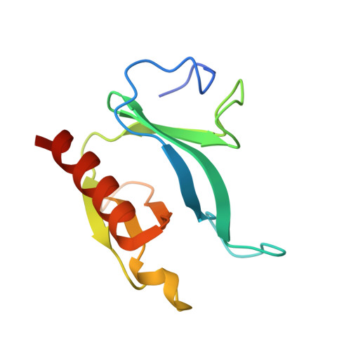
The Skap-hom dimerization and PH domains comprise a 3'-phosphoinositide-gated molecular switch.

Swanson, K.D.Tang, Y.Ceccarelli, D.F.Poy, F.Sliwa, J.P.Neel, B.G.Eck, M.J.

(2008) Mol Cell 32: 564-575

  • DOI: https://doi.org/10.1016/j.molcel.2008.09.022
  • Primary Citation of Related Structures:  
    1U5G, 2OTX

  • PubMed Abstract: 

    PH domains, by binding to phosphoinositides, often serve as membrane-targeting modules. Using crystallographic, biochemical, and cell biological approaches, we have uncovered a mechanism that the integrin-signaling adaptor Skap-hom uses to mediate cytoskeletal interactions. Skap-hom is a homodimer containing an N-terminal four-helix bundle dimerization domain, against which its two PH domains pack in a conformation incompatible with phosphoinositide binding. The isolated PH domains bind PI[3,4,5]P(3), and mutations targeting the dimerization domain or the PH domain's PI[3,4,5]P(3)-binding pocket prevent Skap-hom localization to ruffles. Targeting is retained when the PH domain is deleted or by combined mutation of the PI[3,4,5]P(3)-binding pocket and the PH/dimerization domain interface. Thus, the dimerization and PH domain form a PI[3,4,5]P(3)-responsive molecular switch that controls Skap-hom function.


  • Organizational Affiliation

    Cancer Biology Program, Division of Hematology-Oncology, Department of Medicine, Beth Israel Deaconess Medical Center, Boston, MA 02115, USA.


Macromolecules
Find similar proteins by:  (by identity cutoff)  |  3D Structure
Entity ID: 1
MoleculeChains Sequence LengthOrganismDetailsImage
Src-associated adaptor protein
A, B, C, D
122Mus musculusMutation(s): 2 
Gene Names: SCAP2
UniProt
Find proteins for Q3UND0 (Mus musculus)
Explore Q3UND0 
Go to UniProtKB:  Q3UND0
Entity Groups  
Sequence Clusters30% Identity50% Identity70% Identity90% Identity95% Identity100% Identity
UniProt GroupQ3UND0
Sequence Annotations
Expand
  • Reference Sequence
Experimental Data & Validation

Experimental Data

  • Method: X-RAY DIFFRACTION
  • Resolution: 2.10 Å
  • R-Value Free: 0.271 
  • R-Value Work: 0.213 
  • R-Value Observed: 0.216 
  • Space Group: P 21 21 21
Unit Cell:
Length ( Å )Angle ( ˚ )
a = 59.613α = 90
b = 65.695β = 90
c = 143.767γ = 90
Software Package:
Software NamePurpose
MAR345data collection
SCALEPACKdata scaling
AMoREphasing
REFMACrefinement

Structure Validation

View Full Validation Report


Entry History 

Deposition Data

Revision History  (Full details and data files)

  • Version 1.0: 2005-07-26
    Type: Initial release
  • Version 1.1: 2008-04-30
    Changes: Version format compliance
  • Version 1.2: 2011-07-13
    Changes: Version format compliance
  • Version 1.3: 2017-10-11
    Changes: Refinement description
  • Version 1.4: 2021-10-20
    Changes: Database references
  • Version 1.5: 2023-08-23
    Changes: Data collection, Refinement description