1TEW

STRUCTURE OF HEXAGONAL TURKEY EGG WHITE LYSOZYME AT 1.65 ANGSTROMS RESOLUTION


Experimental Data Snapshot

  • Method: X-RAY DIFFRACTION
  • Resolution: 1.65 Å
  • R-Value Work: 0.206 
  • R-Value Observed: 0.206 

wwPDB Validation   3D Report Full Report


This is version 1.2 of the entry. See complete history


Literature

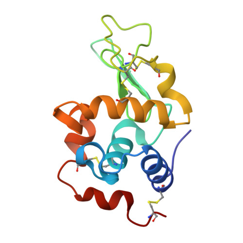
Structure of hexagonal turkey egg-white lysozyme at 1.65A resolution.

Howell, P.L.

(1995) Acta Crystallogr D Biol Crystallogr 51: 654-662

  • DOI: https://doi.org/10.1107/S0907444994013612
  • Primary Citation of Related Structures:  
    1TEW

  • PubMed Abstract: 

    The structure of hexagonal turkey egg-white lysozyme (TEWL) has been determined and refined at 1.65 A resolution. The crystals were grown from a 150 mM potassium thiocyanate solution at pH 4.5 and belong to space group P6(1)22 with unit-cell dimensions a = b = 70.96, c = 83.01 A alpha = beta = 90, gamma = 120 degrees. The crystals were isomorphous with those of hexagonal pH 8.0 TEWL. The coordinates of PDB entry code 3LZ2 were therefore used as the initial model and subjected to rigid-body refinement, simulated annealing and least-squares refinement to a final residual of 0.20. The root-mean-square deviations from the ideal bond distances and angles were 0.016 A and 2.2 degrees, respectively. During the refinement, 86 water molecules and one thiocyanate ion were located in the structure. The thiocyanate ion lies close to the interface between two symmetry-related molecules. The S atom of the ion forms two direct intermolecular contacts with Argl4 and interacts indirectly via a network of water molecules to Arg5 of a symmetry-related molecule. The structure provides direct evidence for the mode of thiocyanate binding to arginine residues and suggests a possible mechanism for the efficiency of thiocyanate in crystallizing basic proteins.


  • Organizational Affiliation

    Division of Biochemistry Research, Hospital for Sick Children, Toronto, Canada.


Macromolecules
Find similar proteins by:  (by identity cutoff)  |  3D Structure
Entity ID: 1
MoleculeChains Sequence LengthOrganismDetailsImage
TURKEY EGG WHITE LYSOZYME129Meleagris gallopavoMutation(s): 0 
EC: 3.2.1.17
UniProt
Find proteins for P00703 (Meleagris gallopavo)
Explore P00703 
Go to UniProtKB:  P00703
Entity Groups  
Sequence Clusters30% Identity50% Identity70% Identity90% Identity95% Identity100% Identity
UniProt GroupP00703
Sequence Annotations
Expand
  • Reference Sequence
Small Molecules
Ligands 1 Unique
IDChains Name / Formula / InChI Key2D Diagram3D Interactions
SCN
Query on SCN

Download Ideal Coordinates CCD File 
B [auth A]THIOCYANATE ION
C N S
ZMZDMBWJUHKJPS-UHFFFAOYSA-M
Experimental Data & Validation

Experimental Data

  • Method: X-RAY DIFFRACTION
  • Resolution: 1.65 Å
  • R-Value Work: 0.206 
  • R-Value Observed: 0.206 
  • Space Group: P 61 2 2
Unit Cell:
Length ( Å )Angle ( ˚ )
a = 70.96α = 90
b = 70.96β = 90
c = 83.01γ = 120
Software Package:
Software NamePurpose
X-PLORmodel building
X-PLORrefinement
PROFFTrefinement
X-PLORphasing

Structure Validation

View Full Validation Report


Entry History 

Deposition Data

  • Released Date: 1995-01-26 
  • Deposition Author(s): Howell, P.L.

Revision History  (Full details and data files)

  • Version 1.0: 1995-01-26
    Type: Initial release
  • Version 1.1: 2008-03-03
    Changes: Version format compliance
  • Version 1.2: 2011-07-13
    Changes: Version format compliance