1T1L

Crystal structure of the long-chain fatty acid transporter FadL


Experimental Data Snapshot

  • Method: X-RAY DIFFRACTION
  • Resolution: 2.80 Å
  • R-Value Free: 0.331 
  • R-Value Work: 0.298 
  • R-Value Observed: 0.306 

wwPDB Validation   3D Report Full Report


This is version 1.3 of the entry. See complete history


Literature

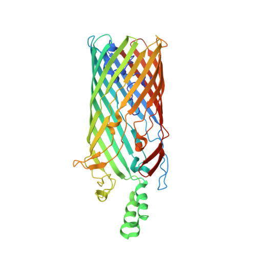
Crystal structure of the long-chain fatty acid transporter FadL.

van den Berg, B.Black, P.N.Clemons Jr., W.M.Rapoport, T.A.

(2004) Science 304: 1506-1509

  • DOI: https://doi.org/10.1126/science.1097524
  • Primary Citation of Related Structures:  
    1T16, 1T1L

  • PubMed Abstract: 

    The mechanisms by which hydrophobic molecules, such as long-chain fatty acids, enter cells are poorly understood. In Gram-negative bacteria, the lipopolysaccharide layer in the outer membrane is an efficient barrier for fatty acids and aromatic hydrocarbons destined for biodegradation. We report crystal structures of the long-chain fatty acid transporter FadL from Escherichia coli at 2.6 and 2.8 angstrom resolution. FadL forms a 14-stranded beta barrel that is occluded by a central hatch domain. The structures suggest that hydrophobic compounds bind to multiple sites in FadL and use a transport mechanism that involves spontaneous conformational changes in the hatch.


  • Organizational Affiliation

    Howard Hughes Medical Institute and Department of Cell Biology, Harvard Medical School, 240 Longwood Avenue, Boston, MA 02115, USA. lvandenberg@hms.harvard.edu


Macromolecules
Find similar proteins by:  (by identity cutoff)  |  3D Structure
Entity ID: 1
MoleculeChains Sequence LengthOrganismDetailsImage
Long-chain fatty acid transport protein
A, B
427Escherichia coliMutation(s): 0 
Gene Names: FADLTTRB2344
Membrane Entity: Yes 
UniProt
Find proteins for P10384 (Escherichia coli (strain K12))
Explore P10384 
Go to UniProtKB:  P10384
Entity Groups  
Sequence Clusters30% Identity50% Identity70% Identity90% Identity95% Identity100% Identity
UniProt GroupP10384
Sequence Annotations
Expand
  • Reference Sequence
Small Molecules
Experimental Data & Validation

Experimental Data

  • Method: X-RAY DIFFRACTION
  • Resolution: 2.80 Å
  • R-Value Free: 0.331 
  • R-Value Work: 0.298 
  • R-Value Observed: 0.306 
  • Space Group: P 31 2 1
Unit Cell:
Length ( Å )Angle ( ˚ )
a = 122.02α = 90
b = 122.02β = 90
c = 164.736γ = 120
Software Package:
Software NamePurpose
CNSrefinement
HKL-2000data reduction
SCALEPACKdata scaling
SOLVEphasing

Structure Validation

View Full Validation Report


Entry History 

Deposition Data

Revision History  (Full details and data files)

  • Version 1.0: 2004-06-15
    Type: Initial release
  • Version 1.1: 2008-04-30
    Changes: Version format compliance
  • Version 1.2: 2011-07-13
    Changes: Version format compliance
  • Version 1.3: 2024-02-14
    Changes: Data collection, Database references, Derived calculations