1T0O

The structure of alpha-galactosidase from Trichoderma reesei complexed with beta-D-galactose


Experimental Data Snapshot

  • Method: X-RAY DIFFRACTION
  • Resolution: 1.96 Å
  • R-Value Free: 0.205 
  • R-Value Work: 0.151 
  • R-Value Observed: 0.157 

wwPDB Validation   3D Report Full Report


Ligand Structure Quality Assessment 


This is version 2.1 of the entry. See complete history


Literature

Crystal structure of alpha-galactosidase from Trichoderma reesei and its complex with galactose: implications for catalytic mechanism

Golubev, A.M.Nagem, R.A.P.Brandao Neto, J.R.Neustroev, K.N.Eneyskaya, E.V.Kulminskaya, A.A.Shabalin, K.A.Savel'ev, A.N.Polikarpov, I.

(2004) J Mol Biol 339: 413-422

  • DOI: https://doi.org/10.1016/j.jmb.2004.03.062
  • Primary Citation of Related Structures:  
    1SZN, 1T0O

  • PubMed Abstract: 

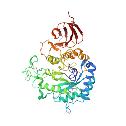
    The crystal structures of alpha-galactosidase from the mesophilic fungus Trichoderma reesei and its complex with the competitive inhibitor, beta-d-galactose, have been determined at 1.54 A and 2.0 A resolution, respectively. The alpha-galactosidase structure was solved by the quick cryo-soaking method using a single Cs derivative. The refined crystallographic model of the alpha-galactosidase consists of two domains, an N-terminal catalytic domain of the (beta/alpha)8 barrel topology and a C-terminal domain which is formed by an antiparallel beta-structure. The protein contains four N-glycosylation sites located in the catalytic domain. Some of the oligosaccharides were found to participate in inter-domain contacts. The galactose molecule binds to the active site pocket located in the center of the barrel of the catalytic domain. Analysis of the alpha-galactosidase- galactose complex reveals the residues of the active site and offers a structural basis for identification of the putative mechanism of the enzymatic reaction. The structure of the alpha-galactosidase closely resembles those of the glycoside hydrolase family 27. The conservation of two catalytic Asp residues, identified for this family, is consistent with a double-displacement reaction mechanism for the alpha-galactosidase. Modeling of possible substrates into the active site reveals specific hydrogen bonds and hydrophobic interactions that could explain peculiarities of the enzyme kinetics.


  • Organizational Affiliation

    Petersburg Nuclear Physics Institute, Gatchina, St Petersburg, 188300, Russia.


Macromolecules
Find similar proteins by:  (by identity cutoff)  |  3D Structure
Entity ID: 1
MoleculeChains Sequence LengthOrganismDetailsImage
alpha-galactosidase417Trichoderma reeseiMutation(s): 0 
EC: 3.2.1.22
UniProt
Find proteins for Q92456 (Hypocrea jecorina)
Explore Q92456 
Go to UniProtKB:  Q92456
Entity Groups  
Sequence Clusters30% Identity50% Identity70% Identity90% Identity95% Identity100% Identity
UniProt GroupQ92456
Sequence Annotations
Expand
  • Reference Sequence
Oligosaccharides

Help

Entity ID: 2
MoleculeChains Length2D Diagram Glycosylation3D Interactions
2-acetamido-2-deoxy-beta-D-glucopyranose-(1-4)-2-acetamido-2-deoxy-beta-D-glucopyranose
B, E
2N-Glycosylation
Glycosylation Resources
GlyTouCan:  G42666HT
GlyCosmos:  G42666HT
GlyGen:  G42666HT
Entity ID: 3
MoleculeChains Length2D Diagram Glycosylation3D Interactions
alpha-D-mannopyranose-(1-3)-[alpha-D-mannopyranose-(1-6)]alpha-D-mannopyranose-(1-6)-[alpha-D-mannopyranose-(1-3)]beta-D-mannopyranose-(1-4)-2-acetamido-2-deoxy-beta-D-glucopyranose-(1-4)-2-acetamido-2-deoxy-beta-D-glucopyranose
C
7N-Glycosylation
Glycosylation Resources
GlyTouCan:  G55220VL
GlyCosmos:  G55220VL
GlyGen:  G55220VL
Entity ID: 4
MoleculeChains Length2D Diagram Glycosylation3D Interactions
alpha-D-mannopyranose-(1-3)-beta-D-mannopyranose-(1-4)-2-acetamido-2-deoxy-beta-D-glucopyranose-(1-4)-2-acetamido-2-deoxy-beta-D-glucopyranose
D
4N-Glycosylation
Glycosylation Resources
GlyTouCan:  G81315DD
GlyCosmos:  G81315DD
GlyGen:  G81315DD
Small Molecules
Ligands 1 Unique
IDChains Name / Formula / InChI Key2D Diagram3D Interactions
GAL
Query on GAL

Download Ideal Coordinates CCD File 
F [auth A]beta-D-galactopyranose
C6 H12 O6
WQZGKKKJIJFFOK-FPRJBGLDSA-N
Experimental Data & Validation

Experimental Data

  • Method: X-RAY DIFFRACTION
  • Resolution: 1.96 Å
  • R-Value Free: 0.205 
  • R-Value Work: 0.151 
  • R-Value Observed: 0.157 
  • Space Group: P 21 21 21
Unit Cell:
Length ( Å )Angle ( ˚ )
a = 57.834α = 90
b = 58.313β = 90
c = 153.66γ = 90
Software Package:
Software NamePurpose
REFMACrefinement
DENZOdata reduction
SCALEPACKdata scaling
AMoREphasing

Structure Validation

View Full Validation Report


Ligand Structure Quality Assessment 


Entry History 

Revision History  (Full details and data files)

  • Version 1.0: 2004-10-12
    Type: Initial release
  • Version 1.1: 2008-04-30
    Changes: Version format compliance
  • Version 1.2: 2011-07-13
    Changes: Non-polymer description, Version format compliance
  • Version 2.0: 2020-07-29
    Type: Remediation
    Reason: Carbohydrate remediation
    Changes: Advisory, Atomic model, Data collection, Derived calculations, Structure summary
  • Version 2.1: 2023-08-23
    Changes: Data collection, Database references, Refinement description, Structure summary