1SZW

Crystal structure of E. coli tRNA pseudouridine synthase TruD


Experimental Data Snapshot

  • Method: X-RAY DIFFRACTION
  • Resolution: 2.00 Å
  • R-Value Free: 0.239 
  • R-Value Work: 0.192 
  • R-Value Observed: 0.194 

wwPDB Validation   3D Report Full Report


This is version 1.3 of the entry. See complete history


Literature

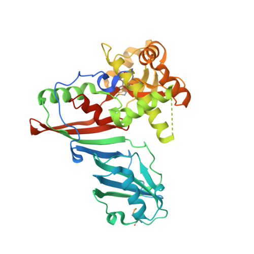
X-ray structure of tRNA pseudouridine synthase TruD reveals an inserted domain with a novel fold

Ericsson, U.B.Nordlund, P.Hallberg, B.M.

(2004) FEBS Lett 565: 59-64

  • DOI: https://doi.org/10.1016/j.febslet.2004.03.085
  • Primary Citation of Related Structures:  
    1SZW

  • PubMed Abstract: 

    Pseudouridine synthases catalyse the isomerisation of uridine to pseudouridine in structural RNA. The pseudouridine synthase TruD, that modifies U13 in tRNA, belongs to a recently identified and large family of pseudouridine synthases present in all kingdoms of life. We report here the crystal structure of Escherichia coli TruD at 2.0 A resolution. The structure reveals an overall V-shaped molecule with an RNA-binding cleft formed between two domains: a catalytic domain and an insertion domain. The catalytic domain has a fold similar to that of the catalytic domains of previously characterised pseudouridine synthases, whereas the insertion domain displays a novel fold.


  • Organizational Affiliation

    Department of Biochemistry and Biophysics, Stockholm University, Roslagstullsbacken 15, SE-114 21 Stockholm, Sweden.


Macromolecules
Find similar proteins by:  (by identity cutoff)  |  3D Structure
Entity ID: 1
MoleculeChains Sequence LengthOrganismDetailsImage
tRNA pseudouridine synthase D
A, B
379Escherichia coliMutation(s): 4 
Gene Names: TRUD
EC: 4.2.1.70
UniProt
Find proteins for Q57261 (Escherichia coli (strain K12))
Explore Q57261 
Go to UniProtKB:  Q57261
Entity Groups  
Sequence Clusters30% Identity50% Identity70% Identity90% Identity95% Identity100% Identity
UniProt GroupQ57261
Sequence Annotations
Expand
  • Reference Sequence
Small Molecules
Modified Residues  1 Unique
IDChains TypeFormula2D DiagramParent
MSE
Query on MSE
A, B
L-PEPTIDE LINKINGC5 H11 N O2 SeMET
Experimental Data & Validation

Experimental Data

  • Method: X-RAY DIFFRACTION
  • Resolution: 2.00 Å
  • R-Value Free: 0.239 
  • R-Value Work: 0.192 
  • R-Value Observed: 0.194 
  • Space Group: P 21 21 21
Unit Cell:
Length ( Å )Angle ( ˚ )
a = 63.18α = 90
b = 108.11β = 90
c = 112.52γ = 90
Software Package:
Software NamePurpose
REFMACrefinement
MAR345data collection
XDSdata scaling
SHELXDphasing

Structure Validation

View Full Validation Report


Entry History 

Deposition Data

Revision History  (Full details and data files)

  • Version 1.0: 2004-04-20
    Type: Initial release
  • Version 1.1: 2008-04-30
    Changes: Version format compliance
  • Version 1.2: 2011-07-13
    Changes: Advisory, Version format compliance
  • Version 1.3: 2017-10-11
    Changes: Refinement description