Crystal structure of a free kappaB DNA: insights into DNA recognition by transcription factor NF-kappaB.
Huang, D.B., Phelps, C.B., Fusco, A.J., Ghosh, G.(2005) J Mol Biol 346: 147-160
- PubMed: 15663934
- DOI: https://doi.org/10.1016/j.jmb.2004.11.042
- Primary Citation of Related Structures:
1SGS - PubMed Abstract:
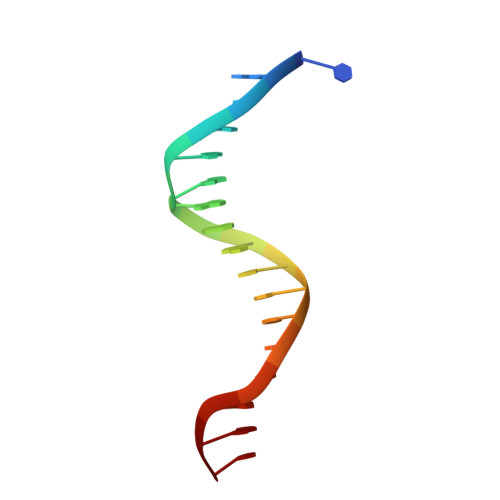
The dimeric NF-kappaB transcription factors regulate gene expression by recognizing specific DNA sequences located within the promoters of target genes. The DNA sequences, referred to as kappaB DNA, are divided into two broad classes. Class I kappaB DNA binds optimally to p50 and p52 NF-kappaB subunits, while class II kappaB DNAs are recognized specifically by the NF-kappaB subunits c-Rel and p65. We determined the X-ray crystal structure of a class II kappaB DNA sequence at 1.60 A resolution. This structure provides a detailed picture of kappaB DNA hydration, counter ion binding, and conformation in the absence of NF-kappaB binding partner. X-ray structures of both class I and class II kappaB DNA bound to NF-kappaB dimers were determined previously. Additionally, the NMR solution structure of a class I kappaB DNA is known. Comparison of the protein-bound and unbound kappaB DNA structures reveals that the free form of both classes approximates ideal B-form DNA more closely. Local geometries about specific DNA bases differ significantly upon binding to NF-kappaB. This is particularly evident at the 5'-GG/CC base-pairs; a signature of NF-kappaB specific DNA binding sequences. Differential phosphate group conformations, minor groove widths, buckle, twist, and tilt angles are observed between bound and unbound kappaB DNA. We observe that the presence of an extra G:C base-pair, 5'- to the GGA sequence in class I kappaB DNA, alters the geometry of the two internal G:C base-pairs within the GGGA tetranucleotide, which explains, at least in part, the structural basis for distinct NF-kappaB dimer recruitment by the two different classes of kappaB DNA. Together, these observations suggest that NF-kappaB dimers recognize specific structural features of kappaB DNA in order to make sequence-specific complexes.
Organizational Affiliation:
Department of Chemistry and Biochemistry, University of California, San Diego, Mail Code 0375, 9500 Gilman Drive, La Jolla, CA 92093-0375, USA.