


Crystal structure of conserved hypothetical protein from Deinicoccus radiodurans
Fedorov, A.A., Fedorov, E.V., Almo, S.C.To be published.
Experimental Data Snapshot
wwPDB Validation 3D Report Full Report

Entity ID: 1 | |||||
|---|---|---|---|---|---|
| Molecule | Chains | Sequence Length | Organism | Details | Image |
| conserved hypothetical protein | 246 | Deinococcus radiodurans | Mutation(s): 0 | 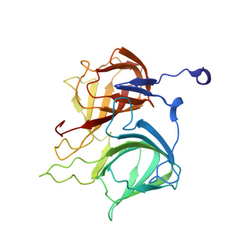 | |
UniProt | |||||
Find proteins for Q9RV77 (Deinococcus radiodurans (strain ATCC 13939 / DSM 20539 / JCM 16871 / CCUG 27074 / LMG 4051 / NBRC 15346 / NCIMB 9279 / VKM B-1422 / R1)) Explore Q9RV77 Go to UniProtKB: Q9RV77 | |||||
Entity Groups | |||||
| Sequence Clusters | 30% Identity50% Identity70% Identity90% Identity95% Identity100% Identity | ||||
| UniProt Group | Q9RV77 | ||||
Sequence AnnotationsExpand | |||||
| |||||
| Length ( Å ) | Angle ( ˚ ) |
|---|---|
| a = 109.37 | α = 90 |
| b = 109.37 | β = 90 |
| c = 95.408 | γ = 90 |
| Software Name | Purpose |
|---|---|
| DENZO | data reduction |
| SCALEPACK | data scaling |
| SOLVE | phasing |
| CNS | refinement |
RCSB PDB (citation) is hosted by
RCSB PDB is a member of the

