1SEH

Crystal structure of E. coli dUTPase complexed with the product dUMP


Experimental Data Snapshot

  • Method: X-RAY DIFFRACTION
  • Resolution: 1.47 Å
  • R-Value Free: 0.184 
  • R-Value Work: 0.158 
  • R-Value Observed: 0.159 

wwPDB Validation   3D Report Full Report


Ligand Structure Quality Assessment 


This is version 1.4 of the entry. See complete history


Literature

Structural Insights into the Catalytic Mechanism of Phosphate Ester Hydrolysis by dUTPase

Barabas, O.Pongracz, V.Kovari, J.Wilmanns, M.Vertessy, B.G.

(2004) J Biol Chem 279: 42907-42915

  • DOI: https://doi.org/10.1074/jbc.M406135200
  • Primary Citation of Related Structures:  
    1RN8, 1RNJ, 1SEH, 1SYL

  • PubMed Abstract: 

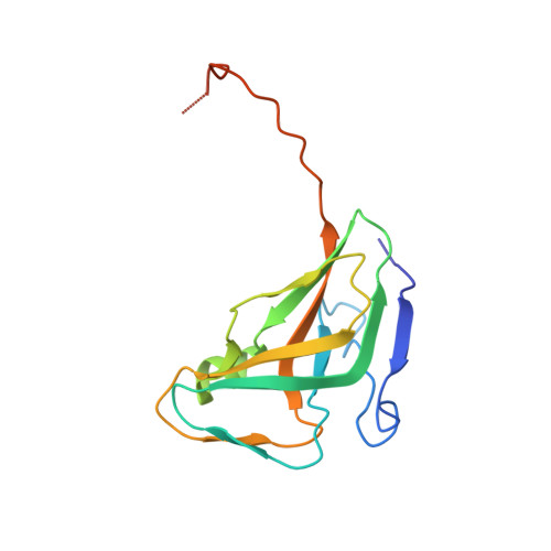
    dUTPase is essential to keep uracil out of DNA. Crystal structures of substrate (dUTP and alpha,beta-imino-dUTP) and product complexes of wild type and mutant dUTPases were determined to reveal how an enzyme responsible for DNA integrity functions. A kinetic analysis of wild type and mutant dUTPases was performed to obtain relevant mechanistic information in solution. Substrate hydrolysis is shown to be initiated via in-line nucleophile attack of a water molecule oriented by an activating conserved aspartate residue. Substrate binding in a catalytically competent conformation is achieved by (i) multiple interactions of the triphosphate moiety with catalysis-assisting Mg2+, (ii) a concerted motion of residues from three conserved enzyme motifs as compared with the apoenzyme, and (iii) an intricate hydrogen-bonding network that includes several water molecules in the active site. Results provide an understanding for the catalytic role of conserved residues in dUTPases.


  • Organizational Affiliation

    Institute of Enzymology, Biological Research Center, Hungarian Academy of Science, Budapest, Karolina út 29-31, H-1113, Hungary.


Macromolecules
Find similar proteins by:  (by identity cutoff)  |  3D Structure
Entity ID: 1
MoleculeChains Sequence LengthOrganismDetailsImage
Deoxyuridine 5'-triphosphate nucleotidohydrolase152Escherichia coliMutation(s): 0 
Gene Names: DUT
EC: 3.6.1.23
UniProt
Find proteins for P06968 (Escherichia coli (strain K12))
Explore P06968 
Go to UniProtKB:  P06968
Entity Groups  
Sequence Clusters30% Identity50% Identity70% Identity90% Identity95% Identity100% Identity
UniProt GroupP06968
Sequence Annotations
Expand
  • Reference Sequence
Small Molecules
Ligands 2 Unique
IDChains Name / Formula / InChI Key2D Diagram3D Interactions
UMP
Query on UMP

Download Ideal Coordinates CCD File 
B [auth A]2'-DEOXYURIDINE 5'-MONOPHOSPHATE
C9 H13 N2 O8 P
JSRLJPSBLDHEIO-SHYZEUOFSA-N
TRS
Query on TRS

Download Ideal Coordinates CCD File 
C [auth A]2-AMINO-2-HYDROXYMETHYL-PROPANE-1,3-DIOL
C4 H12 N O3
LENZDBCJOHFCAS-UHFFFAOYSA-O
Experimental Data & Validation

Experimental Data

  • Method: X-RAY DIFFRACTION
  • Resolution: 1.47 Å
  • R-Value Free: 0.184 
  • R-Value Work: 0.158 
  • R-Value Observed: 0.159 
  • Space Group: P 63 2 2
Unit Cell:
Length ( Å )Angle ( ˚ )
a = 75.257α = 90
b = 75.257β = 90
c = 100.532γ = 120
Software Package:
Software NamePurpose
REFMACrefinement
MAR345data collection
XDSdata scaling
MOLREPphasing

Structure Validation

View Full Validation Report


Ligand Structure Quality Assessment 


Entry History 

Deposition Data

Revision History  (Full details and data files)

  • Version 1.0: 2004-09-07
    Type: Initial release
  • Version 1.1: 2008-04-29
    Changes: Version format compliance
  • Version 1.2: 2011-07-13
    Changes: Derived calculations, Version format compliance
  • Version 1.3: 2017-10-11
    Changes: Refinement description
  • Version 1.4: 2023-10-25
    Changes: Data collection, Database references, Derived calculations, Refinement description