1RHY

Crystal structure of Imidazole Glycerol Phosphate Dehydratase


Experimental Data Snapshot

  • Method: X-RAY DIFFRACTION
  • Resolution: 2.30 Å
  • R-Value Free: 0.228 
  • R-Value Work: 0.189 
  • R-Value Observed: 0.189 

wwPDB Validation   3D Report Full Report


This is version 1.5 of the entry. See complete history


Literature

Crystal structure of imidazole glycerol-phosphate dehydratase: duplication of an unusual fold

Sinha, S.C.Chaudhuri, B.N.Burgner, J.W.Yakovleva, G.Davisson, V.J.Smith, J.L.

(2004) J Biol Chem 279: 15491-15498

  • DOI: https://doi.org/10.1074/jbc.M312733200
  • Primary Citation of Related Structures:  
    1RHY

  • PubMed Abstract: 

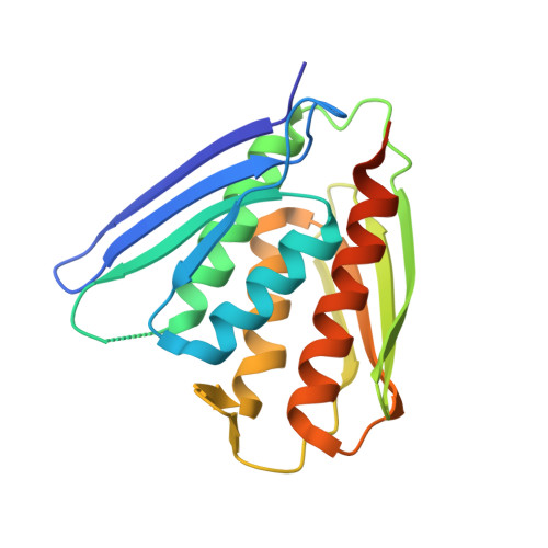
    Imidazole glycerol-phosphate dehydratase (IGPD) catalyzes the sixth step of histidine biosynthesis. The enzyme is of fundamental biochemical interest, because it catalyzes removal of a non-acidic hydrogen atom in the dehydration reaction. It is also a potential target for development of herbicides. IGPD is a metalloenzyme in which transition metals induce aggregation and are required for catalysis. Addition of 1 equivalent of Mn(2+)/subunit is shown by analytical ultracentrifugation to induce the formation of 24-mers from trimeric IGPD. Two histidine-rich motifs may participate in metal binding and aggregation. The 2.3-A crystal structure of metal-free trimeric IGPD from the fungus Filobasidiella neoformans reveals a novel fold containing an internal repeat, apparently the result of gene duplication. The 95-residue alpha/beta half-domain occurs in a few other proteins, including the GHMP kinase superfamily (galacto-homoserine-mevalonate-phosphomevalonate), but duplication to form a compact domain has not been seen elsewhere. Conserved residues cluster at two types of sites in the trimer, each site containing a conserved histidine-rich motif. A model is proposed for the intact, active 24-mer in which all highly conserved residues, including the histidine-rich motifs in both the N- and C-terminal halves of the polypeptide, cluster at a common site between trimers. This site is a candidate for the active site and also for metal binding leading to aggregation of trimers. The structure provides a basis for further studies of enzyme function and mechanism and for development of more potent and specific herbicides.


  • Organizational Affiliation

    Department of Biological Sciences, Purdue University, West Lafayette, Indiana 47907, USA.


Macromolecules
Find similar proteins by:  (by identity cutoff)  |  3D Structure
Entity ID: 1
MoleculeChains Sequence LengthOrganismDetailsImage
Imidazole glycerol phosphate dehydratase
A, B
202Cryptococcus neoformansMutation(s): 0 
Gene Names: HIS3
EC: 4.2.1.19
UniProt
Find proteins for P0CO22 (Cryptococcus neoformans var. neoformans serotype D (strain JEC21 / ATCC MYA-565))
Explore P0CO22 
Go to UniProtKB:  P0CO22
Entity Groups  
Sequence Clusters30% Identity50% Identity70% Identity90% Identity95% Identity100% Identity
UniProt GroupP0CO22
Sequence Annotations
Expand
  • Reference Sequence
Small Molecules
Ligands 5 Unique
IDChains Name / Formula / InChI Key2D Diagram3D Interactions
EMC
Query on EMC

Download Ideal Coordinates CCD File 
D [auth A],
E [auth A],
K [auth B],
L [auth B]
ETHYL MERCURY ION
C2 H5 Hg
MJOUBOKSWBMNGQ-UHFFFAOYSA-N
HG
Query on HG

Download Ideal Coordinates CCD File 
C [auth A],
J [auth B]
MERCURY (II) ION
Hg
BQPIGGFYSBELGY-UHFFFAOYSA-N
SO4
Query on SO4

Download Ideal Coordinates CCD File 
F [auth A],
G [auth A],
H [auth A],
M [auth B],
N [auth B]
SULFATE ION
O4 S
QAOWNCQODCNURD-UHFFFAOYSA-L
GOL
Query on GOL

Download Ideal Coordinates CCD File 
AA [auth B]
I [auth A]
V [auth B]
W [auth B]
X [auth B]
AA [auth B],
I [auth A],
V [auth B],
W [auth B],
X [auth B],
Y [auth B],
Z [auth B]
GLYCEROL
C3 H8 O3
PEDCQBHIVMGVHV-UHFFFAOYSA-N
ACY
Query on ACY

Download Ideal Coordinates CCD File 
O [auth B]
P [auth B]
Q [auth B]
R [auth B]
S [auth B]
O [auth B],
P [auth B],
Q [auth B],
R [auth B],
S [auth B],
T [auth B],
U [auth B]
ACETIC ACID
C2 H4 O2
QTBSBXVTEAMEQO-UHFFFAOYSA-N
Experimental Data & Validation

Experimental Data

  • Method: X-RAY DIFFRACTION
  • Resolution: 2.30 Å
  • R-Value Free: 0.228 
  • R-Value Work: 0.189 
  • R-Value Observed: 0.189 
  • Space Group: P 21 3
Unit Cell:
Length ( Å )Angle ( ˚ )
a = 105.275α = 90
b = 105.275β = 90
c = 105.275γ = 90
Software Package:
Software NamePurpose
XDSdata scaling
SCALEPACKdata scaling
SOLVEphasing
DMmodel building
CNSrefinement
XDSdata reduction
DMphasing

Structure Validation

View Full Validation Report


Entry History 

Deposition Data

Revision History  (Full details and data files)

  • Version 1.0: 2004-05-04
    Type: Initial release
  • Version 1.1: 2008-04-29
    Changes: Version format compliance
  • Version 1.2: 2011-07-13
    Changes: Derived calculations, Version format compliance
  • Version 1.3: 2011-11-16
    Changes: Atomic model
  • Version 1.4: 2017-10-11
    Changes: Refinement description
  • Version 1.5: 2024-02-14
    Changes: Data collection, Database references, Derived calculations