1QAT

1-PHOSPHATIDYLINOSITOL-4,5-BISPHOSPHATE PHOSPHODIESTERASE DELTA COMPLEX WITH SAMARIUM (III) CHLORIDE


Experimental Data Snapshot

  • Method: X-RAY DIFFRACTION
  • Resolution: 3.00 Å
  • R-Value Free: 0.261 
  • R-Value Work: 0.194 
  • R-Value Observed: 0.194 

wwPDB Validation   3D Report Full Report


This is version 1.4 of the entry. See complete history


Literature

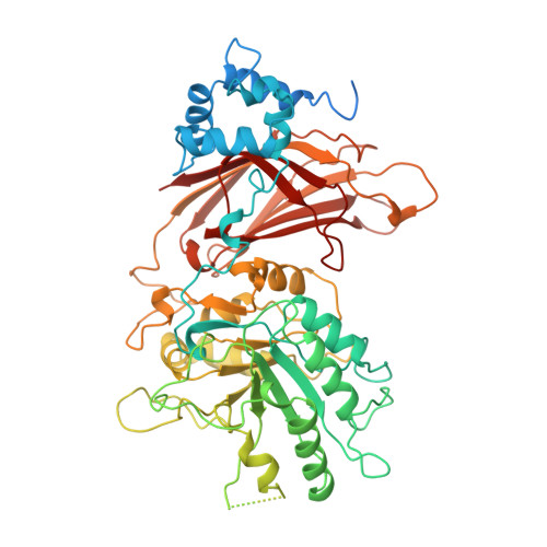
C2 domain conformational changes in phospholipase C-delta 1.

Grobler, J.A.Essen, L.O.Williams, R.L.Hurley, J.H.

(1996) Nat Struct Biol 3: 788-795

  • DOI: https://doi.org/10.1038/nsb0996-788
  • Primary Citation of Related Structures:  
    1QAS, 1QAT

  • PubMed Abstract: 

    The structure of the PH-domain truncated core of rat phosphoinositide-specific phospholipase C-delta 1 has been determined at 2.4 A resolution and compared to the structure previously determined in a different crystal form. The stereochemical relationship between the EF, catalytic, and C2 domains is essentially identical. The Ca2+ analogue Sm3+ binds at two sites between the jaws of the C2 domain. Sm3+ binding ejects three lysine residues which bridge the gap between the jaws and occupy the Ca2+ site in the apoenzyme, triggering a conformational change in the jaws. The distal sections of the C2 jaws move apart, opening the mouth by 9 A and creating a gap large enough to bind a phospholipid headgroup.


  • Organizational Affiliation

    Laboratory of Molecular Biology, National Institute of Diabetes, Digestive, and Kidney Diseases, National Institutes of Health, Bethesda, Maryland 20892-0580, USA.


Macromolecules
Find similar proteins by:  (by identity cutoff)  |  3D Structure
Entity ID: 1
MoleculeChains Sequence LengthOrganismDetailsImage
PHOSPHOLIPASE C DELTA-1
A, B
622Rattus norvegicusMutation(s): 0 
EC: 3.1.4.11
UniProt
Find proteins for P10688 (Rattus norvegicus)
Explore P10688 
Go to UniProtKB:  P10688
Entity Groups  
Sequence Clusters30% Identity50% Identity70% Identity90% Identity95% Identity100% Identity
UniProt GroupP10688
Sequence Annotations
Expand
  • Reference Sequence
Experimental Data & Validation

Experimental Data

  • Method: X-RAY DIFFRACTION
  • Resolution: 3.00 Å
  • R-Value Free: 0.261 
  • R-Value Work: 0.194 
  • R-Value Observed: 0.194 
  • Space Group: P 1
Unit Cell:
Length ( Å )Angle ( ˚ )
a = 61.6α = 66.4
b = 75.1β = 85.6
c = 86.4γ = 89.8
Software Package:
Software NamePurpose
X-PLORmodel building
X-PLORrefinement
DENZOdata reduction
SCALEPACKdata scaling
X-PLORphasing

Structure Validation

View Full Validation Report


Entry History 

Deposition Data

Revision History  (Full details and data files)

  • Version 1.0: 1997-02-12
    Type: Initial release
  • Version 1.1: 2008-03-24
    Changes: Version format compliance
  • Version 1.2: 2011-07-13
    Changes: Version format compliance
  • Version 1.3: 2024-02-14
    Changes: Data collection, Database references, Derived calculations, Other
  • Version 1.4: 2024-04-03
    Changes: Refinement description