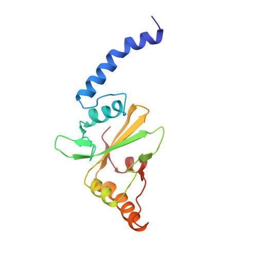
1PVU

THE CRYSTAL STRUCTURE OF PVUII ENDONUCLEASE REVEALS EXTENSIVE STRUCTURAL HOMOLOGIES TO ECORV


Experimental Data Snapshot

  • Method: X-RAY DIFFRACTION
  • Resolution: 2.40 Å
  • R-Value Work: 0.184 
  • R-Value Observed: 0.184 

wwPDB Validation   3D Report Full Report


This is version 1.4 of the entry. See complete history


Literature

Crystal structure of PvuII endonuclease reveals extensive structural homologies to EcoRV.

Athanasiadis, A.Vlassi, M.Kotsifaki, D.Tucker, P.A.Wilson, K.S.Kokkinidis, M.

(1994) Nat Struct Biol 1: 469-475

  • DOI: https://doi.org/10.1038/nsb0794-469
  • Primary Citation of Related Structures:  
    1PVU

  • PubMed Abstract: 

    The crystal structure of the dimeric PvuII restriction endonuclease (R.PvuII) has been determined at a resolution of 2.4A. The protein has a mixed alpha/beta architecture and consists of two subdomains. Despite a lack of sequence homology, extensive structural similarities exist between one R.PvuII subdomain and the DNA-binding subdomain of EcoRV endonuclease (R.EcoRV); the dimerization subdomains are unrelated. Within the similar domains, flexible segments of R.PvuII are topologically equivalent to the DNA-binding turns of R.EcoRV; potential catalytic residues can be deduced from the structural similarities to R.EcoRV. Conformational flexibility is important for the interaction with DNA. A possible classification of endonuclease structures on the basis of the positions of the scissile phosphates is discussed.


  • Organizational Affiliation

    Department of Biology, University of Crete, Greece.


Macromolecules
Find similar proteins by:  (by identity cutoff)  |  3D Structure
Entity ID: 1
MoleculeChains Sequence LengthOrganismDetailsImage
Pvu II
A, B
156Proteus vulgarisMutation(s): 0 
EC: 3.1.21.4
UniProt
Find proteins for P23657 (Proteus hauseri)
Explore P23657 
Go to UniProtKB:  P23657
Entity Groups  
Sequence Clusters30% Identity50% Identity70% Identity90% Identity95% Identity100% Identity
UniProt GroupP23657
Sequence Annotations
Expand
  • Reference Sequence
Experimental Data & Validation

Experimental Data

  • Method: X-RAY DIFFRACTION
  • Resolution: 2.40 Å
  • R-Value Work: 0.184 
  • R-Value Observed: 0.184 
  • Space Group: P 21 21 2
Unit Cell:
Length ( Å )Angle ( ˚ )
a = 84.65α = 90
b = 106.46β = 90
c = 47γ = 90
Software Package:
Software NamePurpose
X-PLORmodel building
X-PLORrefinement
X-PLORphasing

Structure Validation

View Full Validation Report


Entry History 

Deposition Data

Revision History  (Full details and data files)

  • Version 1.0: 1995-03-31
    Type: Initial release
  • Version 1.1: 2008-03-24
    Changes: Version format compliance
  • Version 1.2: 2011-07-13
    Changes: Version format compliance
  • Version 1.3: 2017-11-29
    Changes: Derived calculations, Other
  • Version 1.4: 2024-02-14
    Changes: Data collection, Database references