1PV7

Crystal structure of lactose permease with TDG


Experimental Data Snapshot

  • Method: X-RAY DIFFRACTION
  • Resolution: 3.60 Å
  • R-Value Free: 0.296 
  • R-Value Work: 0.271 
  • R-Value Observed: 0.271 

wwPDB Validation   3D Report Full Report


This is version 3.1 of the entry. See complete history


Literature

Structure and mechanism of the lactose permease of Escherichia coli

Abramson, J.Smirnova, I.Kasho, V.Verner, G.Kaback, H.R.Iwata, S.

(2003) Science 301: 610-615

  • DOI: https://doi.org/10.1126/science.1088196
  • Primary Citation of Related Structures:  
    1PV6, 1PV7

  • PubMed Abstract: 

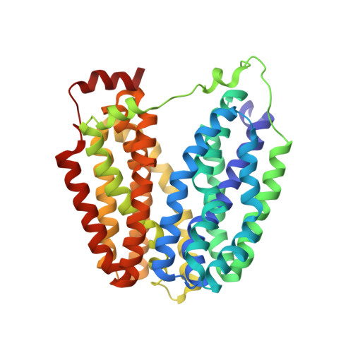
    Membrane transport proteins that transduce free energy stored in electrochemical ion gradients into a concentration gradient are a major class of membrane proteins. We report the crystal structure at 3.5 angstroms of the Escherichia coli lactose permease, an intensively studied member of the major facilitator superfamily of transporters. The molecule is composed of N- and C-terminal domains, each with six transmembrane helices, symmetrically positioned within the permease. A large internal hydrophilic cavity open to the cytoplasmic side represents the inward-facing conformation of the transporter. The structure with a bound lactose homolog, beta-D-galactopyranosyl-1-thio-beta-D-galactopyranoside, reveals the sugar-binding site in the cavity, and residues that play major roles in substrate recognition and proton translocation are identified. We propose a possible mechanism for lactose/proton symport (co-transport) consistent with both the structure and a large body of experimental data.


  • Organizational Affiliation

    Department of Biological Sciences, Imperial College London, London SW7 2AZ, UK.


Macromolecules
Find similar proteins by:  (by identity cutoff)  |  3D Structure
Entity ID: 1
MoleculeChains Sequence LengthOrganismDetailsImage
Lactose permease
A, B
417Escherichia coliMutation(s): 1 
Membrane Entity: Yes 
UniProt
Find proteins for P02920 (Escherichia coli (strain K12))
Explore P02920 
Go to UniProtKB:  P02920
Entity Groups  
Sequence Clusters30% Identity50% Identity70% Identity90% Identity95% Identity100% Identity
UniProt GroupP02920
Sequence Annotations
Expand
  • Reference Sequence
Oligosaccharides

Help

Entity ID: 2
MoleculeChains Length2D Diagram Glycosylation3D Interactions
beta-D-galactopyranose-(1-1)-1-thio-beta-D-galactopyranoseC [auth E],
D [auth F]
2N/AN/A
Glycosylation Resources
GlyTouCan:  G61532BD
GlyCosmos:  G61532BD
Biologically Interesting Molecules (External Reference) 1 Unique
Experimental Data & Validation

Experimental Data

  • Method: X-RAY DIFFRACTION
  • Resolution: 3.60 Å
  • R-Value Free: 0.296 
  • R-Value Work: 0.271 
  • R-Value Observed: 0.271 
  • Space Group: P 21 21 21
Unit Cell:
Length ( Å )Angle ( ˚ )
a = 101.351α = 90
b = 125.845β = 90
c = 188.118γ = 90
Software Package:
Software NamePurpose
DENZOdata reduction
SCALEPACKdata scaling
SHARPphasing
CNSrefinement

Structure Validation

View Full Validation Report


Entry History 

Deposition Data

Revision History  (Full details and data files)

  • Version 1.0: 2003-08-12
    Type: Initial release
  • Version 1.1: 2008-04-29
    Changes: Version format compliance
  • Version 1.2: 2011-07-13
    Changes: Version format compliance
  • Version 2.0: 2020-07-29
    Type: Remediation
    Reason: Carbohydrate remediation
    Changes: Atomic model, Data collection, Derived calculations, Non-polymer description, Structure summary
  • Version 3.0: 2020-08-12
    Changes: Atomic model, Data collection, Derived calculations, Structure summary
  • Version 3.1: 2021-11-10
    Changes: Database references