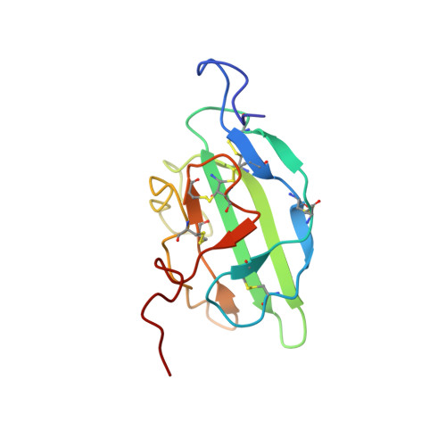
Solution structure and backbone dynamics of the TGF-beta type II receptor extracellular domain
Deep, S., Walker III, K.P., Shu, Z., Hinck, A.P.(2003) Biochemistry 42: 10126-10139
- PubMed: 12939140
- DOI: https://doi.org/10.1021/bi034366a
- Primary Citation of Related Structures:
1PLO - PubMed Abstract:
Isoforms of transforming growth factor beta (TGFbeta) are 25 kDa homodimeric polypeptides that signal by binding and bringing together two related, functionally distinct cell surface receptors designated as TbetaR1 and TbetaR2. Here, we report the solution structure of the 13.8 kDa extracellular domain of human TbetaR2 (ecTbetaR2) as calculated from N(N)-H(N), C(alpha)-H(alpha), and C(alpha)-C(O) residual dipolar coupling restraints in conjunction with NOE distance, dihedral angle, and scalar coupling restraints. Comparison of the free ecTbetaR2 solution structure with the TGFbeta3-bound ecTbetaR2 crystal structure reveals backbone conformations that superimpose with RMSDs of 1.0 A over the regions of regular secondary structure and 1.4 A overall. The differences in structure fall mainly in loop regions that are either poorly defined by the available NMR data or are involved in crystal contacts. The noted similarities between the NMR structure of the free form and the crystal structure of the TGFbeta-bound form are also consistent with the close correspondence, 0.16 A RMSD for regions of secondary structure and 0.51 A RMSD overall, for the crystal structure of free ecTbetaR2 as compared to the crystal structure of TGFbeta3-bound ecTbetaR2. Despite the apparent similarities between the free and the bound forms, there appears to be small but significant differences in structure involving the interfacial contact region of the receptor. Measurements of backbone (15)N relaxation times and interpretation of these by the model-free formalism with axial diffusional anisotropy further reveal significant ms to micros time scale motions centered about two of the conserved disulfide bonds and in several residues that comprise the TGFbeta binding surface. Together, these observations indicate that binding likely occurs through a mechanism with a small component of induced fit character, whereby flexibility within the receptor facilitates the transition to the TGFbeta-bound state.
- Department of Biochemistry, University of Texas Health Science Center at San Antonio, San Antonio, Texas 78229-3900, USA.
Organizational Affiliation: