


The 2 A crystal structure of protein paaC from E. Coli
Zhang, R., Joachimiak, A., Edwards, A., Savchenko, A., Skarina, T.To be published.
Experimental Data Snapshot
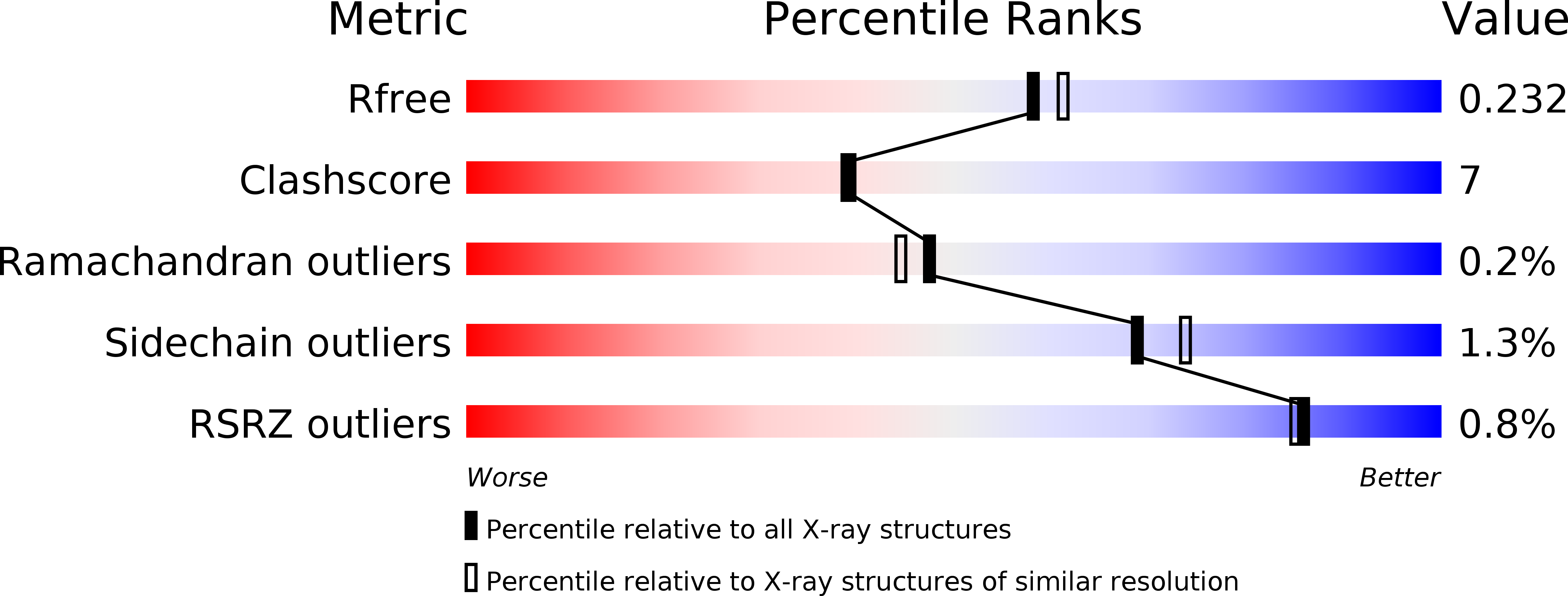
wwPDB Validation 3D Report Full Report
Entity ID: 1 | |||||
|---|---|---|---|---|---|
| Molecule | Chains | Sequence Length | Organism | Details | Image |
| Phenylacetic acid degradation protein paaC | 249 | Escherichia coli K-12 | Mutation(s): 0 Gene Names: PAAC OR B1390 | 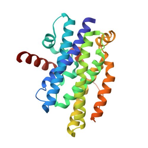 | |
UniProt | |||||
Find proteins for P76079 (Escherichia coli (strain K12)) Explore P76079 Go to UniProtKB: P76079 | |||||
Entity Groups | |||||
| Sequence Clusters | 30% Identity50% Identity70% Identity90% Identity95% Identity100% Identity | ||||
| UniProt Group | P76079 | ||||
Sequence AnnotationsExpand | |||||
| |||||
| Length ( Å ) | Angle ( ˚ ) |
|---|---|
| a = 59.216 | α = 90 |
| b = 95.707 | β = 90 |
| c = 127.088 | γ = 90 |
| Software Name | Purpose |
|---|---|
| CNS | refinement |
| d*TREK | data reduction |
| HKL-2000 | data scaling |
| CNS | phasing |
RCSB PDB (citation) is hosted by
RCSB PDB is a member of the

