1OKQ

LAMININ ALPHA 2 CHAIN LG4-5 DOMAIN PAIR, CA1 SITE MUTANT


Experimental Data Snapshot

  • Method: X-RAY DIFFRACTION
  • Resolution: 2.80 Å
  • R-Value Free: 0.270 
  • R-Value Work: 0.220 
  • R-Value Observed: 0.220 

wwPDB Validation   3D Report Full Report


This is version 1.4 of the entry. See complete history


Literature

Distinct Requirements for Heparin and Alpha-Dystroglycan Binding Revealed by Structure-Based Mutagenesis of the Laminin Alpha2 Lg4-Lg5 Domain Pair

Wizemann, H.Garbe, J.H.O.Friedrich, M.V.K.Timpl, R.Sasaki, T.Hohenester, E.

(2003) J Mol Biol 332: 635

  • DOI: https://doi.org/10.1016/s0022-2836(03)00848-9
  • Primary Citation of Related Structures:  
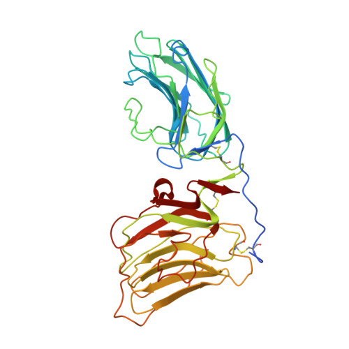
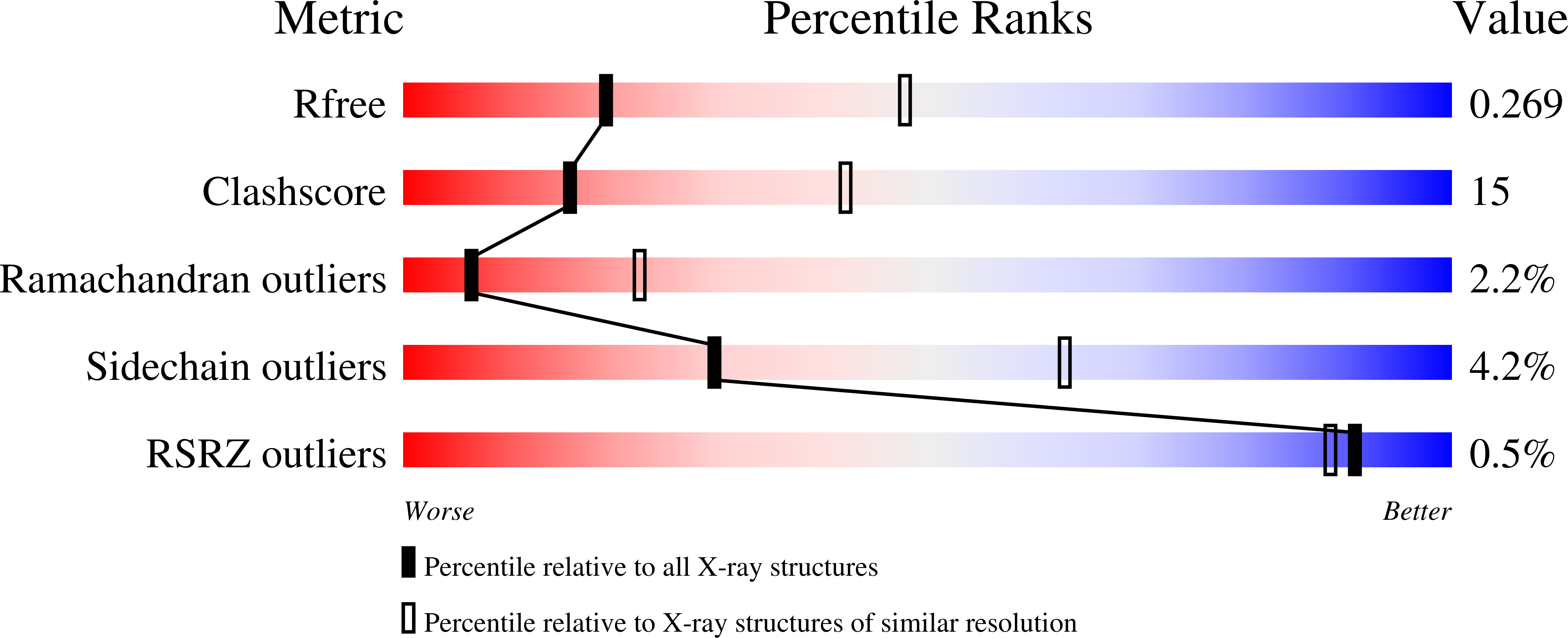
    1OKQ

  • PubMed Abstract: 

    Laminin-2 (alpha2beta1gamma1) is found in basement membranes surrounding muscle and peripheral nerve cells. Several types of cellular receptors bind to the laminin G-like (LG) domains at the C terminus of the alpha2 chain, the interaction with alpha-dystroglycan (alpha-DG) being particularly important in muscle. We have used site-directed mutagenesis and in vitro binding assays to map the binding sites on the laminin alpha2 chain LG4-LG5 domain pair for alpha-DG, heparin and sulfatides. Calcium-dependent alpha-DG recognition requires the calcium ion in LG4, but not the one in LG5, as well as basic residues in both LG domains. Heparin and sulfatides also bind to basic residues in both LG domains, but there is little overlap in the binding sites for alpha-DG and heparin/sulfatides. The results should prove useful for the molecular dissection of laminin-receptor interactions in vivo.


  • Organizational Affiliation

    Max-Planck-Institut für Biochemie, D-82152, Martinsried, Germany.


Macromolecules
Find similar proteins by:  (by identity cutoff)  |  3D Structure
Entity ID: 1
MoleculeChains Sequence LengthOrganismDetailsImage
LAMININ ALPHA 2 CHAIN394Mus musculusMutation(s): 2 
UniProt
Find proteins for Q60675 (Mus musculus)
Explore Q60675 
Go to UniProtKB:  Q60675
Entity Groups  
Sequence Clusters30% Identity50% Identity70% Identity90% Identity95% Identity100% Identity
UniProt GroupQ60675
Sequence Annotations
Expand
  • Reference Sequence
Small Molecules
Ligands 1 Unique
IDChains Name / Formula / InChI Key2D Diagram3D Interactions
CA
Query on CA

Download Ideal Coordinates CCD File 
B [auth A]CALCIUM ION
Ca
BHPQYMZQTOCNFJ-UHFFFAOYSA-N
Experimental Data & Validation

Experimental Data

  • Method: X-RAY DIFFRACTION
  • Resolution: 2.80 Å
  • R-Value Free: 0.270 
  • R-Value Work: 0.220 
  • R-Value Observed: 0.220 
  • Space Group: C 2 2 21
Unit Cell:
Length ( Å )Angle ( ˚ )
a = 70.11α = 90
b = 110.734β = 90
c = 123.835γ = 90
Software Package:
Software NamePurpose
CNSrefinement
MOSFLMdata reduction
SCALAdata scaling
AMoREphasing

Structure Validation

View Full Validation Report


Entry History 

Deposition Data

Revision History  (Full details and data files)

  • Version 1.0: 2003-09-11
    Type: Initial release
  • Version 1.1: 2011-05-08
    Changes: Version format compliance
  • Version 1.2: 2011-07-13
    Changes: Version format compliance
  • Version 1.3: 2019-05-22
    Changes: Data collection, Other, Refinement description
  • Version 1.4: 2023-12-13
    Changes: Data collection, Database references, Derived calculations, Other, Refinement description