1OJH

Crystal structure of NblA from PCC 7120


Experimental Data Snapshot

  • Method: X-RAY DIFFRACTION
  • Resolution: 1.80 Å
  • R-Value Free: 0.217 
  • R-Value Work: 0.182 
  • R-Value Observed: 0.184 

wwPDB Validation   3D Report Full Report


This is version 1.2 of the entry. See complete history


Literature

Crystal Structure of Nbla from Anabaena Sp. Pcc 7120, a Small Protein Playing a Key Role in Phycobilisome Degradation.

Bienert, R.Baier, K.Volkmer, R.Lockau, W.Heinemann, U.

(2006) J Biol Chem 281: 5216

  • DOI: https://doi.org/10.1074/jbc.M507243200
  • Primary Citation of Related Structures:  
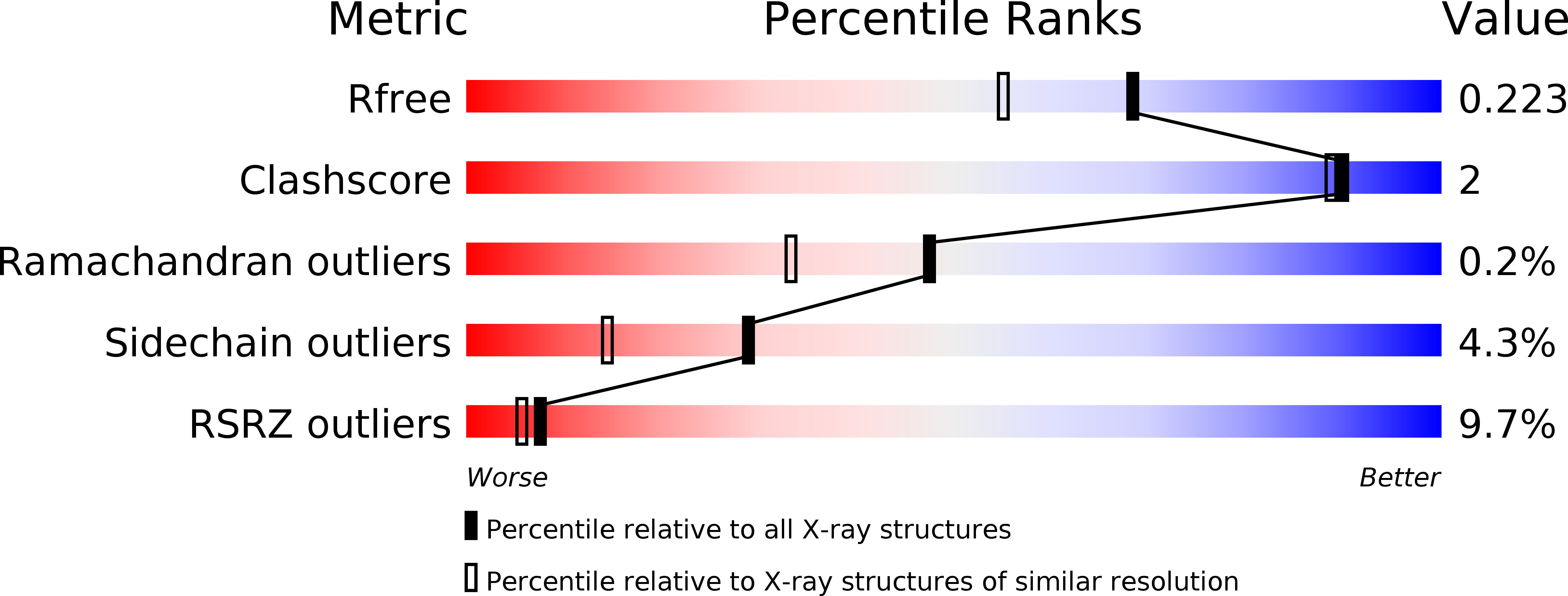
    1OJH

  • PubMed Abstract: 

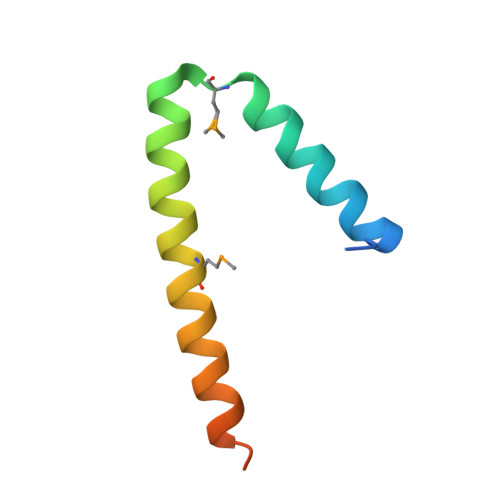
    Cyanobacterial light-harvesting complexes, the phycobilisomes, are proteolytically degraded when the organisms are starved for combined nitrogen, a process referred to as chlorosis or bleaching. Gene nblA, present in all phycobilisome-containing organisms, encodes a protein of about 7 kDa that plays a key role in phycobilisome degradation. The mode of action of NblA in this degradation process is poorly understood. Here we presented the 1.8-A crystal structure of NblA from Anabaena sp. PCC 7120. In the crystal, NblA is present as a four-helix bundle formed by dimers, the basic structural units. By using pull-down assays with immobilized NblA and peptide scanning, we showed that NblA specifically binds to the alpha-subunits of phycocyanin and phycoerythrocyanin, the main building blocks of the phycobilisome rod structure. By site-directed mutagenesis, we identified amino acid residues in NblA that are involved in phycobilisome binding. The results provided evidence that NblA is directly involved in phycobilisome degradation, and the results allowed us to present a model that gives insight into the interaction of this small protein with the phycobilisomes.


  • Organizational Affiliation

    Crystallography Group, Max-Delbrück Center for Molecular Medicine, D-13125 Berlin.


Macromolecules
Find similar proteins by:  (by identity cutoff)  |  3D Structure
Entity ID: 1
MoleculeChains Sequence LengthOrganismDetailsImage
NBLA
A, B, C, D, E
A, B, C, D, E, F, G, H, I, J, K, L
65Nostoc sp. PCC 7120 = FACHB-418Mutation(s): 0 
UniProt
Find proteins for Q8YNP7 (Nostoc sp. (strain PCC 7120 / SAG 25.82 / UTEX 2576))
Explore Q8YNP7 
Go to UniProtKB:  Q8YNP7
Entity Groups  
Sequence Clusters30% Identity50% Identity70% Identity90% Identity95% Identity100% Identity
UniProt GroupQ8YNP7
Sequence Annotations
Expand
  • Reference Sequence
Small Molecules
Modified Residues  1 Unique
IDChains TypeFormula2D DiagramParent
MSE
Query on MSE
A, B, C, D, E
A, B, C, D, E, F, G, H, I, J, K, L
L-PEPTIDE LINKINGC5 H11 N O2 SeMET
Experimental Data & Validation

Experimental Data

  • Method: X-RAY DIFFRACTION
  • Resolution: 1.80 Å
  • R-Value Free: 0.217 
  • R-Value Work: 0.182 
  • R-Value Observed: 0.184 
  • Space Group: P 1 21 1
Unit Cell:
Length ( Å )Angle ( ˚ )
a = 43.176α = 90
b = 95.918β = 97.05
c = 104.835γ = 90
Software Package:
Software NamePurpose
REFMACrefinement
XDSdata reduction
XSCALEdata scaling
SOLVEphasing

Structure Validation

View Full Validation Report


Entry History 

Deposition Data

Revision History  (Full details and data files)

  • Version 1.0: 2004-07-15
    Type: Initial release
  • Version 1.1: 2011-07-13
    Changes: Advisory, Version format compliance
  • Version 1.2: 2011-08-03
    Changes: Database references, Derived calculations, Non-polymer description, Other, Refinement description, Structure summary