1NST

THE SULFOTRANSFERASE DOMAIN OF HUMAN HAPARIN SULFATE N-DEACETYLASE/N-SULFOTRANSFERASE


Experimental Data Snapshot

  • Method: X-RAY DIFFRACTION
  • Resolution: 2.30 Å
  • R-Value Free: 0.257 
  • R-Value Work: 0.210 
  • R-Value Observed: 0.210 

wwPDB Validation   3D Report Full Report


This is version 1.4 of the entry. See complete history


Literature

Crystal structure of the sulfotransferase domain of human heparan sulfate N-deacetylase/ N-sulfotransferase 1.

Kakuta, Y.Sueyoshi, T.Negishi, M.Pedersen, L.C.

(1999) J Biol Chem 274: 10673-10676

  • DOI: https://doi.org/10.1074/jbc.274.16.10673
  • Primary Citation of Related Structures:  
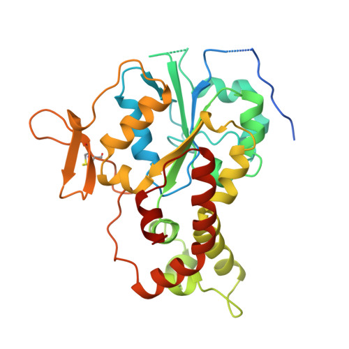
    1NST

  • PubMed Abstract: 

    Heparan sulfate N-deacetylase/N-sulfotransferase (HSNST) catalyzes the first and obligatory step in the biosynthesis of heparan sulfates and heparin. The crystal structure of the sulfotransferase domain (NST1) of human HSNST-1 has been determined at 2.3-A resolution in a binary complex with 3'-phosphoadenosine 5'-phosphate (PAP). NST1 is approximately spherical with an open cleft, and consists of a single alpha/beta fold with a central five-stranded parallel beta-sheet and a three-stranded anti-parallel beta-sheet bearing an interstrand disulfide bond. The structural regions alpha1, alpha6, beta1, beta7, 5'-phosphosulfate binding loop (between beta1 and alpha1), and a random coil (between beta8 and alpha13) constitute the PAP binding site of NST1. The alpha6 and random coil (between beta2 and alpha2), which form an open cleft near the 5'-phosphate of the PAP molecule, may provide interactions for substrate binding. The conserved residue Lys-614 is in position to form a hydrogen bond with the bridge oxygen of the 5'-phosphate.


  • Organizational Affiliation

    Pharmacogenetics Section, Laboratory of Reproductive and Developmental Toxicology, NIEHS, National Institutes of Health, Research Triangle Park, North Carolina 27709, USA.


Macromolecules
Find similar proteins by:  (by identity cutoff)  |  3D Structure
Entity ID: 1
MoleculeChains Sequence LengthOrganismDetailsImage
HEPARAN SULFATE N-DEACETYLASE/N-SULFOTRANSFERASE325Homo sapiensMutation(s): 0 
EC: 2.8.2
UniProt & NIH Common Fund Data Resources
Find proteins for P52848 (Homo sapiens)
Explore P52848 
Go to UniProtKB:  P52848
PHAROS:  P52848
GTEx:  ENSG00000070614 
Entity Groups  
Sequence Clusters30% Identity50% Identity70% Identity90% Identity95% Identity100% Identity
UniProt GroupP52848
Sequence Annotations
Expand
  • Reference Sequence
Small Molecules
Ligands 1 Unique
IDChains Name / Formula / InChI Key2D Diagram3D Interactions
A3P
Query on A3P

Download Ideal Coordinates CCD File 
B [auth A]ADENOSINE-3'-5'-DIPHOSPHATE
C10 H15 N5 O10 P2
WHTCPDAXWFLDIH-KQYNXXCUSA-N
Experimental Data & Validation

Experimental Data

  • Method: X-RAY DIFFRACTION
  • Resolution: 2.30 Å
  • R-Value Free: 0.257 
  • R-Value Work: 0.210 
  • R-Value Observed: 0.210 
  • Space Group: P 1 21 1
Unit Cell:
Length ( Å )Angle ( ˚ )
a = 45.417α = 90
b = 54.501β = 100.05
c = 68.938γ = 90
Software Package:
Software NamePurpose
X-PLORrefinement
SCALEPACKdata scaling
SHELX-96model building
X-PLORmodel building
SHELX-96refinement
DENZOdata reduction
SHELX-96phasing
X-PLORphasing

Structure Validation

View Full Validation Report


Entry History 

Deposition Data

Revision History  (Full details and data files)

  • Version 1.0: 1999-09-16
    Type: Initial release
  • Version 1.1: 2008-03-24
    Changes: Version format compliance
  • Version 1.2: 2011-07-13
    Changes: Version format compliance
  • Version 1.3: 2019-07-17
    Changes: Data collection, Refinement description
  • Version 1.4: 2019-08-14
    Changes: Data collection