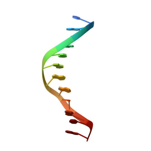
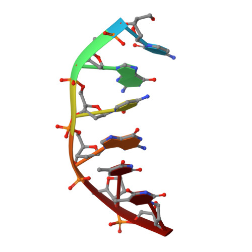
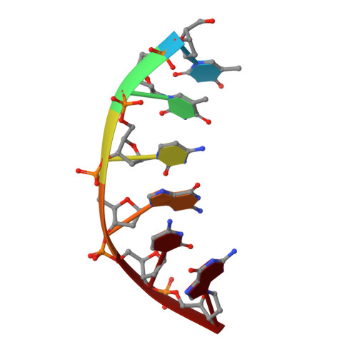
1NDN

MOLECULAR STRUCTURE OF NICKED DNA. MODEL T4


Experimental Data Snapshot

  • Method: X-RAY DIFFRACTION
  • Resolution: 3.00 Å
  • R-Value Observed: 0.186 

wwPDB Validation   3D Report Full Report


This is version 1.3 of the entry. See complete history


Literature

Molecular structure of nicked DNA: a substrate for DNA repair enzymes.

Aymami, J.Coll, M.van der Marel, G.A.van Boom, J.H.Wang, A.H.Rich, A.

(1990) Proc Natl Acad Sci U S A 87: 2526-2530

  • DOI: https://doi.org/10.1073/pnas.87.7.2526
  • Primary Citation of Related Structures:  
    1NDN, 1VTE

  • PubMed Abstract: 

    The molecular structure of a nicked dodecamer DNA double helix, made of a ternary system containing d(CGCGAAAACGCG) + d(CGCGTT) + d(TTCGCG) oligonucleotides, has been determined by x-ray diffraction analysis at 3 A resolution. The molecule adopts a B-DNA conformation, not unlike those found in intact dodecamer DNA molecules crystallized in a somewhat different crystal lattice, despite a gap due to the absence of a phosphate group in the molecule. The helix has a distinct narrow minor groove near the center of the molecule at the AAAA region. This suggests that the internal stabilizing forces due to base stacking and hydrogen-bonding interactions are sufficient to overcome the loss of connectivity associated with the disruption of the covalent backbone of DNA.


  • Organizational Affiliation

    Department of Biology, Massachusetts Institute of Technology, Cambridge 02139.


Macromolecules

Find similar nucleic acids by:  Sequence   |   3D Structure  

Entity ID: 1
MoleculeChains LengthOrganismImage
DNA (5'-D(*CP*GP*CP*GP*AP*AP*AP*AP*CP*GP*CP*G)-3')12N/A
Sequence Annotations
Expand
  • Reference Sequence

Find similar nucleic acids by:  Sequence   |   3D Structure  

Entity ID: 2
MoleculeChains LengthOrganismImage
DNA (5'-D(*CP*GP*CP*GP*TP*T)-3')6N/A
Sequence Annotations
Expand
  • Reference Sequence

Find similar nucleic acids by:  Sequence   |   3D Structure  

Entity ID: 3
MoleculeChains LengthOrganismImage
DNA (5'-D(*TP*TP*CP*GP*CP*G)-3')6N/A
Sequence Annotations
Expand
  • Reference Sequence
Experimental Data & Validation

Experimental Data

  • Method: X-RAY DIFFRACTION
  • Resolution: 3.00 Å
  • R-Value Observed: 0.186 
  • Space Group: P 21 21 21
Unit Cell:
Length ( Å )Angle ( ˚ )
a = 25.99α = 90
b = 44.03β = 90
c = 66.62γ = 90
Software Package:
Software NamePurpose
ULTIMAmodel building
NUCLSQrefinement
ULTIMAphasing

Structure Validation

View Full Validation Report


Entry History 

Deposition Data

Revision History  (Full details and data files)

  • Version 1.0: 1992-07-14
    Type: Initial release
  • Version 1.1: 2008-05-22
    Changes: Version format compliance
  • Version 1.2: 2011-07-13
    Changes: Version format compliance
  • Version 1.3: 2024-02-14
    Changes: Data collection, Database references