1M33

Crystal Structure of BioH at 1.7 A


Experimental Data Snapshot

  • Method: X-RAY DIFFRACTION
  • Resolution: 1.70 Å
  • R-Value Free: 0.188 
  • R-Value Work: 0.145 
  • R-Value Observed: 0.147 

wwPDB Validation   3D Report Full Report


This is version 1.4 of the entry. See complete history


Literature

Integrating structure, bioinformatics, and enzymology to discover function: BioH, a new carboxylesterase from Escherichia coli.

Sanishvili, R.Yakunin, A.F.Laskowski, R.A.Skarina, T.Evdokimova, E.Doherty-Kirby, A.Lajoie, G.A.Thornton, J.M.Arrowsmith, C.H.Savchenko, A.Joachimiak, A.Edwards, A.M.

(2003) J Biol Chem 278: 26039-26045

  • DOI: https://doi.org/10.1074/jbc.M303867200
  • Primary Citation of Related Structures:  
    1M33

  • PubMed Abstract: 

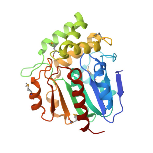
    Structural proteomics projects are generating three-dimensional structures of novel, uncharacterized proteins at an increasing rate. However, structure alone is often insufficient to deduce the specific biochemical function of a protein. Here we determined the function for a protein using a strategy that integrates structural and bioinformatics data with parallel experimental screening for enzymatic activity. BioH is involved in biotin biosynthesis in Escherichia coli and had no previously known biochemical function. The crystal structure of BioH was determined at 1.7 A resolution. An automated procedure was used to compare the structure of BioH with structural templates from a variety of different enzyme active sites. This screen identified a catalytic triad (Ser82, His235, and Asp207) with a configuration similar to that of the catalytic triad of hydrolases. Analysis of BioH with a panel of hydrolase assays revealed a carboxylesterase activity with a preference for short acyl chain substrates. The combined use of structural bioinformatics with experimental screens for detecting enzyme activity could greatly enhance the rate at which function is determined from structure.


  • Organizational Affiliation

    Biosciences Division, Argonne National Laboratory, Argonne, Illinois, 60439, USA.


Macromolecules
Find similar proteins by:  (by identity cutoff)  |  3D Structure
Entity ID: 1
MoleculeChains Sequence LengthOrganismDetailsImage
BioH protein258Escherichia coliMutation(s): 6 
Gene Names: BioH
UniProt
Find proteins for P13001 (Escherichia coli (strain K12))
Explore P13001 
Go to UniProtKB:  P13001
Entity Groups  
Sequence Clusters30% Identity50% Identity70% Identity90% Identity95% Identity100% Identity
UniProt GroupP13001
Sequence Annotations
Expand
  • Reference Sequence
Small Molecules
Modified Residues  1 Unique
IDChains TypeFormula2D DiagramParent
MSE
Query on MSE
A
L-PEPTIDE LINKINGC5 H11 N O2 SeMET
Experimental Data & Validation

Experimental Data

  • Method: X-RAY DIFFRACTION
  • Resolution: 1.70 Å
  • R-Value Free: 0.188 
  • R-Value Work: 0.145 
  • R-Value Observed: 0.147 
  • Space Group: P 43
Unit Cell:
Length ( Å )Angle ( ˚ )
a = 75.21α = 90
b = 75.21β = 90
c = 49.256γ = 90
Software Package:
Software NamePurpose
REFMACrefinement
dtDisplaydata collection
HKL-2000data reduction
CNSrefinement
d*TREKdata scaling
d*TREKdata reduction
DTDISPLAYdata reduction
HKL-2000data scaling
CNSphasing

Structure Validation

View Full Validation Report


Entry History 

Revision History  (Full details and data files)

  • Version 1.0: 2003-01-21
    Type: Initial release
  • Version 1.1: 2008-04-28
    Changes: Version format compliance
  • Version 1.2: 2011-07-13
    Changes: Advisory, Version format compliance
  • Version 1.3: 2017-10-11
    Changes: Advisory, Refinement description
  • Version 1.4: 2019-07-24
    Changes: Data collection, Derived calculations, Refinement description