1LRR

CRYSTAL STRUCTURE OF E. COLI SEQA COMPLEXED WITH HEMIMETHYLATED DNA


Experimental Data Snapshot

  • Method: X-RAY DIFFRACTION
  • Resolution: 2.65 Å
  • R-Value Free: 0.278 
  • R-Value Work: 0.248 

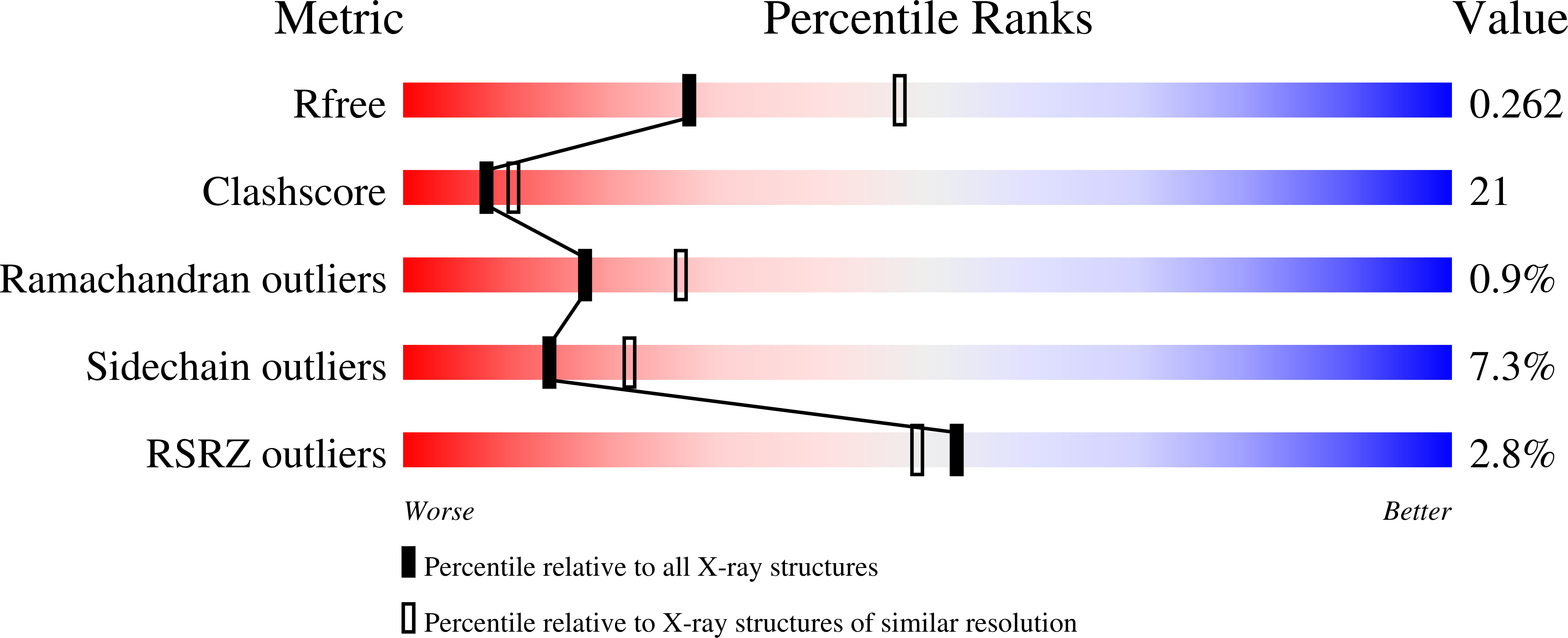
wwPDB Validation   3D Report Full Report


This is version 1.5 of the entry. See complete history


Literature

Insights into negative modulation of E. coli replication initiation from the structure of SeqA-hemimethylated DNA complex

Guarne, A.Zhao, Q.Guirlando, R.Yang, W.

(2002) Nat Struct Biol 9: 839-843

  • DOI: https://doi.org/10.1038/nsb857
  • Primary Citation of Related Structures:  
    1LRR

  • PubMed Abstract: 

    The SeqA protein binds clusters of fully methylated or hemimethylated GATC sequences at oriC and negatively modulates the initiation of DNA replication. We find that SeqA can be proteolytically cleaved into an N-terminal multimerization and a C-terminal DNA-binding domain and have determined the crystal structure of the C-terminal domain in complex with a hemimethylated GATC site. SeqA makes direct hydrogen bonds and van der Waals contacts with the hemimethylated A-T base pair in addition to interactions with the surrounding bases and DNA backbone. The tetrameric protein-DNA complex found in the crystal suggests that SeqA binds multiple GATC sites on separate DNA duplexes, altering the overall DNA topology and sequestering oriC from replication initiation.


  • Organizational Affiliation

    Laboratory of Molecular Biology, National Institute of Diabetes and Digestive and Kidney Diseases, National Institutes of Health, Bethesda, Maryland 20892, USA.


Macromolecules

Find similar proteins by:  (by identity cutoff)  |  3D Structure
Entity ID: 3
MoleculeChains Sequence LengthOrganismDetailsImage
SeqA proteinE [auth A],
F [auth D]
131Escherichia coliMutation(s): 0 
Gene Names: seqA
UniProt
Find proteins for P0AFY8 (Escherichia coli (strain K12))
Explore P0AFY8 
Go to UniProtKB:  P0AFY8
Entity Groups  
Sequence Clusters30% Identity50% Identity70% Identity90% Identity95% Identity100% Identity
UniProt GroupP0AFY8
Sequence Annotations
Expand
  • Reference Sequence

Find similar nucleic acids by:  Sequence   |   3D Structure  

Entity ID: 1
MoleculeChains LengthOrganismImage
5'-D(*CP*AP*CP*CP*GP*AP*TP*CP*GP*AP*CP*T)-3'A [auth B],
C [auth E]
12N/A
Sequence Annotations
Expand
  • Reference Sequence

Find similar nucleic acids by:  Sequence   |   3D Structure  

Entity ID: 2
MoleculeChains LengthOrganismImage
5'-D(*AP*GP*TP*CP*GP*(6MA)P*TP*CP*GP*GP*TP*G)-3'B [auth C],
D [auth F]
12N/A
Sequence Annotations
Expand
  • Reference Sequence
Experimental Data & Validation

Experimental Data

  • Method: X-RAY DIFFRACTION
  • Resolution: 2.65 Å
  • R-Value Free: 0.278 
  • R-Value Work: 0.248 
  • Space Group: P 42
Unit Cell:
Length ( Å )Angle ( ˚ )
a = 106.493α = 90
b = 106.493β = 90
c = 49.078γ = 90
Software Package:
Software NamePurpose
CNSrefinement
DENZOdata reduction
SCALEPACKdata scaling
CNSphasing

Structure Validation

View Full Validation Report


Entry History 

Deposition Data

Revision History  (Full details and data files)

  • Version 1.0: 2002-12-11
    Type: Initial release
  • Version 1.1: 2008-04-28
    Changes: Version format compliance
  • Version 1.2: 2011-07-13
    Changes: Version format compliance
  • Version 1.3: 2013-05-29
    Changes: Atomic model
  • Version 1.4: 2024-02-14
    Changes: Data collection, Database references, Derived calculations
  • Version 1.5: 2024-04-03
    Changes: Refinement description