1L2Q

Crystal Structure of the Methanosarcina barkeri Monomethylamine Methyltransferase (MtmB)


Experimental Data Snapshot

  • Method: X-RAY DIFFRACTION
  • Resolution: 1.70 Å
  • R-Value Free: 0.175 
  • R-Value Work: 0.161 

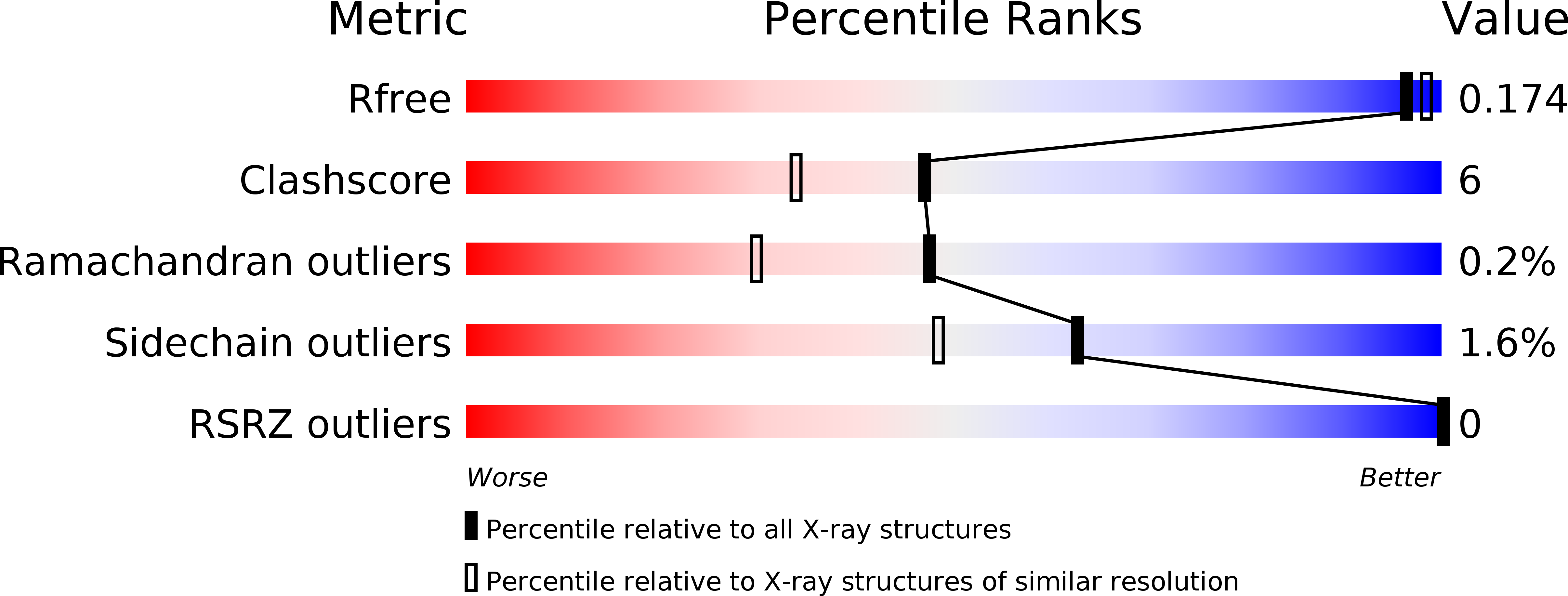
wwPDB Validation   3D Report Full Report


This is version 1.3 of the entry. See complete history


Literature

A new UAG-encoded residue in the structure of a methanogen methyltransferase.

Hao, B.Gong, W.Ferguson, T.K.James, C.M.Krzycki, J.A.Chan, M.K.

(2002) Science 296: 1462-1466

  • DOI: https://doi.org/10.1126/science.1069556
  • Primary Citation of Related Structures:  
    1L2Q, 1NTH

  • PubMed Abstract: 

    Genes encoding methanogenic methylamine methyltransferases all contain an in-frame amber (UAG) codon that is read through during translation. We have identified the UAG-encoded residue in a 1.55 angstrom resolution structure of the Methanosarcina barkeri monomethylamine methyltransferase (MtmB). This structure reveals a homohexamer comprised of individual subunits with a TIM barrel fold. The electron density for the UAG-encoded residue is distinct from any of the 21 natural amino acids. Instead it appears consistent with a lysine in amide-linkage to (4R,5R)-4-substituted-pyrroline-5-carboxylate. We suggest that this amino acid be named l-pyrrolysine.


  • Organizational Affiliation

    Department of Biochemistry, The Ohio State University, 484 West 12th Avenue, Columbus, OH 43210, USA.


Macromolecules
Find similar proteins by:  (by identity cutoff)  |  3D Structure
Entity ID: 1
MoleculeChains Sequence LengthOrganismDetailsImage
monomethylamine methyltransferase458Methanosarcina barkeriMutation(s): 0 
UniProt
Find proteins for O30642 (Methanosarcina barkeri)
Explore O30642 
Go to UniProtKB:  O30642
Entity Groups  
Sequence Clusters30% Identity50% Identity70% Identity90% Identity95% Identity100% Identity
UniProt GroupO30642
Sequence Annotations
Expand
  • Reference Sequence
Small Molecules
Ligands 1 Unique
IDChains Name / Formula / InChI Key2D Diagram3D Interactions
NH4
Query on NH4

Download Ideal Coordinates CCD File 
B [auth A]AMMONIUM ION
H4 N
QGZKDVFQNNGYKY-UHFFFAOYSA-O
Modified Residues  1 Unique
IDChains TypeFormula2D DiagramParent
XPL
Query on XPL
A
L-PEPTIDE LINKINGC12 H24 N4 O3PYL
Experimental Data & Validation

Experimental Data

  • Method: X-RAY DIFFRACTION
  • Resolution: 1.70 Å
  • R-Value Free: 0.175 
  • R-Value Work: 0.161 
  • Space Group: P 63 2 2
Unit Cell:
Length ( Å )Angle ( ˚ )
a = 158.07α = 90
b = 158.07β = 90
c = 135.891γ = 120
Software Package:
Software NamePurpose
PHASESphasing
CNSrefinement
DENZOdata reduction
SCALEPACKdata scaling

Structure Validation

View Full Validation Report


Entry History 

Deposition Data

Revision History  (Full details and data files)

  • Version 1.0: 2002-06-05
    Type: Initial release
  • Version 1.1: 2008-04-28
    Changes: Version format compliance
  • Version 1.2: 2011-07-13
    Changes: Derived calculations, Version format compliance
  • Version 1.3: 2014-08-13
    Changes: Non-polymer description, Structure summary