1KWS

CRYSTAL STRUCTURE OF BETA1,3-GLUCURONYLTRANSFERASE I IN COMPLEX WITH THE ACTIVE UDP-GLCUA DONOR


Experimental Data Snapshot

  • Method: X-RAY DIFFRACTION
  • Resolution: 2.10 Å
  • R-Value Free: 0.223 
  • R-Value Work: 0.187 
  • R-Value Observed: 0.188 

wwPDB Validation   3D Report Full Report


Ligand Structure Quality Assessment 


This is version 1.4 of the entry. See complete history


Literature

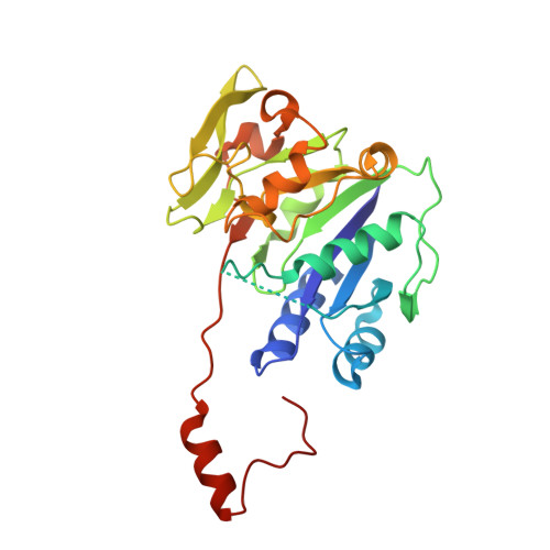
Crystal structure of beta 1,3-glucuronyltransferase I in complex with active donor substrate UDP-GlcUA.

Pedersen, L.C.Darden, T.A.Negishi, M.

(2002) J Biol Chem 277: 21869-21873

  • DOI: https://doi.org/10.1074/jbc.M112343200
  • Primary Citation of Related Structures:  
    1KWS

  • PubMed Abstract: 

    Beta1,3-glucuronyltransferase (GlcAT-I) is an essential enzyme involved in heparan sulfate and chondroitin sulfate biosynthesis. GlcAT-I is an inverting glycosyltransferase that catalyzes the transfer of glucuronic acid (GlcUA) to the common growing linker region Galbeta1-3Galbeta1-4Xyl that is attached to a serine side chain of a core protein. Previously the structure of GlcAT-I has been solved in the presence of the donor product UDP and an acceptor analog Galbeta1-3Galbeta1-4Xyl (Pedersen, L. C., Tsuchida, K., Kitagawa, H., Sugahara, K., Darden, T. A. & Negishi, M. (2000) J. Biol. Chem. 275, 34580-34585). Here we report the x-ray crystal structure of GlcAT-I in complex with the complete donor UDP-GlcUA, thereby providing structures of an inverting glycosyltransferase in which both the complete donor and acceptor substrates are present in the active site. This structure supports the in-line displacement reaction mechanism previously proposed. It also provides information on the essential amino acid residues that determine donor substrate specificity.


  • Organizational Affiliation

    Laboratory of Reproductive and Developmental Toxicology, NIEHS, National Institutes of Health, Research Triangle Park, North Carolina 27709, USA.


Macromolecules
Find similar proteins by:  (by identity cutoff)  |  3D Structure
Entity ID: 1
MoleculeChains Sequence LengthOrganismDetailsImage
BETA-1,3-GLUCURONYLTRANSFERASE 3
A, B
261Homo sapiensMutation(s): 0 
UniProt & NIH Common Fund Data Resources
Find proteins for O94766 (Homo sapiens)
Explore O94766 
Go to UniProtKB:  O94766
PHAROS:  O94766
GTEx:  ENSG00000149541 
Entity Groups  
Sequence Clusters30% Identity50% Identity70% Identity90% Identity95% Identity100% Identity
UniProt GroupO94766
Sequence Annotations
Expand
  • Reference Sequence
Experimental Data & Validation

Experimental Data

  • Method: X-RAY DIFFRACTION
  • Resolution: 2.10 Å
  • R-Value Free: 0.223 
  • R-Value Work: 0.187 
  • R-Value Observed: 0.188 
  • Space Group: P 1 21 1
Unit Cell:
Length ( Å )Angle ( ˚ )
a = 57.625α = 90
b = 47.978β = 93.1
c = 102.152γ = 90
Software Package:
Software NamePurpose
CNSrefinement
SCALEPACKdata scaling
CNSphasing

Structure Validation

View Full Validation Report


Ligand Structure Quality Assessment 


Entry History 

Deposition Data

Revision History  (Full details and data files)

  • Version 1.0: 2002-06-19
    Type: Initial release
  • Version 1.1: 2008-04-27
    Changes: Version format compliance
  • Version 1.2: 2011-07-13
    Changes: Version format compliance
  • Version 1.3: 2017-10-11
    Changes: Refinement description
  • Version 1.4: 2023-08-16
    Changes: Data collection, Database references, Derived calculations, Refinement description