1KHH

Crystal Structure of Guanidinoacetate Methyltransferase from Rat Liver: A Template Structure of Protein Arginine Methyltransferase


Experimental Data Snapshot

  • Method: X-RAY DIFFRACTION
  • Resolution: 2.50 Å
  • R-Value Free: 0.284 
  • R-Value Work: 0.214 
  • R-Value Observed: 0.221 

wwPDB Validation   3D Report Full Report


Ligand Structure Quality Assessment 


This is version 1.4 of the entry. See complete history


Literature

Crystal structure of guanidinoacetate methyltransferase from rat liver: a model structure of protein arginine methyltransferase.

Komoto, J.Huang, Y.Takata, Y.Yamada, T.Konishi, K.Ogawa, H.Gomi, T.Fujioka, M.Takusagawa, F.

(2002) J Mol Biol 320: 223-235

  • DOI: https://doi.org/10.1016/S0022-2836(02)00448-5
  • Primary Citation of Related Structures:  
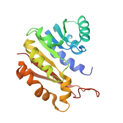
    1KHH

  • PubMed Abstract: 

    Guanidinoacetate methyltransferase (GAMT) is the enzyme that catalyzes the last step of creatine biosynthesis. The enzyme is found in abundance in the livers of all vertebrates. Recombinant rat liver GAMT has been crystallized with S-adenosylhomocysteine (SAH), and the crystal structure has been determined at 2.5 A resolution. The 36 amino acid residues at the N terminus were cleaved during the purification and the truncated enzyme was crystallized. The truncated enzyme forms a dimer, and each subunit contains one SAH molecule in the active site. Arg220 of the partner subunit forms a pair of hydrogen bonds with Asp134 at the guanidinoacetate-binding site. On the basis of the crystal structure, site-directed mutagenesis on Asp134, and chemical modification and limited proteolysis studies, we propose a catalytic mechanism of this enzyme. The truncated GAMT dimer structure can be seen as a ternary complex of protein arginine methyltransferase (one subunit) complexed with a protein substrate (the partner subunit) and the product SAH. Therefore, this structure provides insight into the structure and catalysis of protein arginine methyltransferases.


  • Organizational Affiliation

    Department of Molecular Biosciences, The University of Kansas, 1200 Sunnyside Avenue, 2034 Howorth Hall, Lawrence, KS 66045-7534, USA.


Macromolecules
Find similar proteins by:  (by identity cutoff)  |  3D Structure
Entity ID: 1
MoleculeChains Sequence LengthOrganismDetailsImage
Guanidinoacetate methyltransferase
A, B
198Rattus norvegicusMutation(s): 0 
EC: 2.1.1.2
UniProt
Find proteins for P10868 (Rattus norvegicus)
Explore P10868 
Go to UniProtKB:  P10868
Entity Groups  
Sequence Clusters30% Identity50% Identity70% Identity90% Identity95% Identity100% Identity
UniProt GroupP10868
Sequence Annotations
Expand
  • Reference Sequence
Small Molecules
Experimental Data & Validation

Experimental Data

  • Method: X-RAY DIFFRACTION
  • Resolution: 2.50 Å
  • R-Value Free: 0.284 
  • R-Value Work: 0.214 
  • R-Value Observed: 0.221 
  • Space Group: P 43
Unit Cell:
Length ( Å )Angle ( ˚ )
a = 54.25α = 90
b = 54.25β = 90
c = 156.56γ = 90
Software Package:
Software NamePurpose
SCALEPACKdata scaling
CCP4model building
X-PLORrefinement
CCP4phasing

Structure Validation

View Full Validation Report


Ligand Structure Quality Assessment 


Entry History 

Deposition Data

Revision History  (Full details and data files)

  • Version 1.0: 2001-12-12
    Type: Initial release
  • Version 1.1: 2008-04-27
    Changes: Version format compliance
  • Version 1.2: 2011-07-13
    Changes: Version format compliance
  • Version 1.3: 2017-10-11
    Changes: Refinement description
  • Version 1.4: 2024-02-14
    Changes: Data collection, Database references, Derived calculations