1JT0

Crystal structure of a cooperative QacR-DNA complex


Experimental Data Snapshot

  • Method: X-RAY DIFFRACTION
  • Resolution: 2.90 Å
  • R-Value Free: 0.258 
  • R-Value Work: 0.222 
  • R-Value Observed: 0.222 

wwPDB Validation   3D Report Full Report


This is version 1.4 of the entry. See complete history


Literature

Structural basis for cooperative DNA binding by two dimers of the multidrug-binding protein QacR.

Schumacher, M.A.Miller, M.C.Grkovic, S.Brown, M.H.Skurray, R.A.Brennan, R.G.

(2002) EMBO J 21: 1210-1218

  • DOI: https://doi.org/10.1093/emboj/21.5.1210
  • Primary Citation of Related Structures:  
    1JT0

  • PubMed Abstract: 

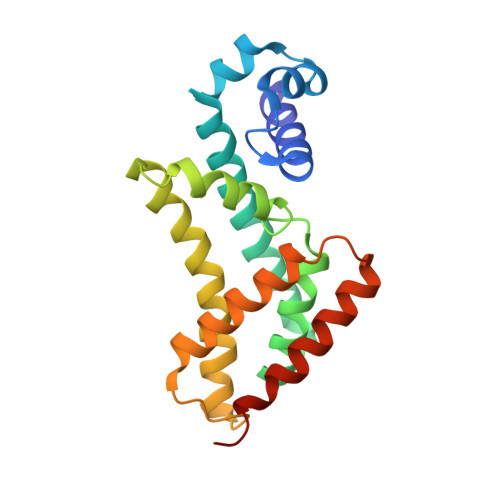
    The Staphylococcus aureus multidrug-binding protein QacR represses transcription of the qacA multidrug transporter gene and is induced by multiple structurally dissimilar drugs. QacR is a member of the TetR/CamR family of transcriptional regulators, which share highly homologous N-terminal DNA-binding domains connected to seemingly non-homologous ligand-binding domains. Unlike other TetR members, which bind approximately 15 bp operators, QacR recognizes an unusually long 28 bp operator, IR1, which it appears to bind cooperatively. To elucidate the DNA-binding mechanism of QacR, we determined the 2.90 A resolution crystal structure of a QacR-IR1 complex. Strikingly, our data reveal that the DNA recognition mode of QacR is distinct from TetR and involves the binding of a pair of QacR dimers. In this unique binding mode, recognition at each IR1 half-site is mediated by a complement of DNA contacts made by two helix-turn-helix motifs. The inferred cooperativity does not arise from cross-dimer protein-protein contacts, but from the global undertwisting and major groove widening elicited by the binding of two QacR dimers.


  • Organizational Affiliation

    Department of Biochemistry and Molecular Biology, Oregon Health & Science University, Portland, OR 97201-3098, USA.


Macromolecules

Find similar proteins by:  (by identity cutoff)  |  3D Structure
Entity ID: 2
MoleculeChains Sequence LengthOrganismDetailsImage
HYPOTHETICAL TRANSCRIPTIONAL REGULATOR IN QACA 5'REGIONC [auth A],
D [auth B],
E [auth C],
F [auth D]
194Staphylococcus aureusMutation(s): 2 
UniProt
Find proteins for P0A0N4 (Staphylococcus aureus)
Explore P0A0N4 
Go to UniProtKB:  P0A0N4
Entity Groups  
Sequence Clusters30% Identity50% Identity70% Identity90% Identity95% Identity100% Identity
UniProt GroupP0A0N4
Sequence Annotations
Expand
  • Reference Sequence
Find similar nucleic acids by:  (by identity cutoff)  |  3D Structure
Entity ID: 1
MoleculeChains LengthOrganismImage
QACA operatorA [auth E],
B [auth F]
28N/A
Sequence Annotations
Expand
  • Reference Sequence
Experimental Data & Validation

Experimental Data

  • Method: X-RAY DIFFRACTION
  • Resolution: 2.90 Å
  • R-Value Free: 0.258 
  • R-Value Work: 0.222 
  • R-Value Observed: 0.222 
  • Space Group: P 65
Unit Cell:
Length ( Å )Angle ( ˚ )
a = 174.7α = 90
b = 174.7β = 90
c = 151.95γ = 120
Software Package:
Software NamePurpose
MOSFLMdata reduction
SCALAdata scaling
CNSrefinement
CCP4data scaling
CNSphasing

Structure Validation

View Full Validation Report


Entry History 

Deposition Data

Revision History  (Full details and data files)

  • Version 1.0: 2002-03-08
    Type: Initial release
  • Version 1.1: 2008-04-27
    Changes: Version format compliance
  • Version 1.2: 2011-07-13
    Changes: Version format compliance
  • Version 1.3: 2021-10-27
    Changes: Database references, Derived calculations
  • Version 1.4: 2024-02-07
    Changes: Data collection