The high-resolution structure of the histidine-containing phosphocarrier protein HPr from Escherichia coli determined by restrained molecular dynamics from nuclear magnetic resonance nuclear Overhauser effect data.
van Nuland, N.A., Hangyi, I.W., van Schaik, R.C., Berendsen, H.J., van Gunsteren, W.F., Scheek, R.M., Robillard, G.T.(1994) J Mol Biol 237: 544-559
- PubMed: 8158637
- DOI: https://doi.org/10.1006/jmbi.1994.1254
- Primary Citation of Related Structures:
1HDN - PubMed Abstract:
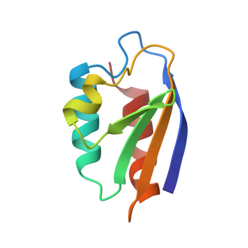
The solution structure of the histidine-containing phosphocarrier protein HPr from Escherichia coli has been determined by NMR in combination with distance geometry and restrained molecular dynamics. The structure is based on 1520 experimental restraints identified from both three-dimensional 1H-1H-13C and 1H-1H-15N nuclear Overhauser effect multiple-quantum coherence spectroscopy and two-dimensional 1H-1H nuclear Overhauser effect spectra. Thirty-two four-dimensional coordinate frames were produced by metric matrix distance geometry, subjected to distance bounds driven dynamics, projected into three-dimensional space and again subjected to distance-bounds driven dynamics. These 32 distance geometry structures were refined further by restrained molecular dynamics (40 ps) in the GROMOS in vacuo force field. All 32 structures reached acceptable energy minima while satisfying the imposed restraints. Two of these structures were subjected to a further 200 ps of molecular dynamics simulation in water, using time-dependent distance restraining, followed by a 200 ps free simulation without any distance restraining. The resulting structure is very similar to the X-ray structure of Bacillus subtilis HPr, but differs mainly in the position of the two loops containing the active site histidine residue 15 and residues 53 to 57 relative to the rest of the structure. The unfavorable phi torsion angle that was found for residue 16 in the active center of unphosphorylated Streptococcus faecalis HPr was proposed to play a role in the activity of the protein. Although present at the early stages of the structure calculations, this torsion-angle strain disappeared in the final model obtained from molecular dynamics simulations in water using time-averaged distance restraining and upon releasing the distance restraints. This suggests that the strain may be an artifact of crystallization conditions instead of an essential element in the phosphorylation/dephosphorylation process.
Organizational Affiliation:
Groningen Biomolecular Sciences and Biotechnology Institute, University of Groningen, The Netherlands.