1HBI

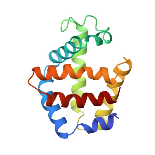
CRYSTAL STRUCTURE OF OXYGENATED SCAPHARCA DIMERIC HEMOGLOBIN AT 1.7 ANGSTROMS RESOLUTION


Experimental Data Snapshot

  • Method: X-RAY DIFFRACTION
  • Resolution: 1.70 Å
  • R-Value Observed: 0.157 

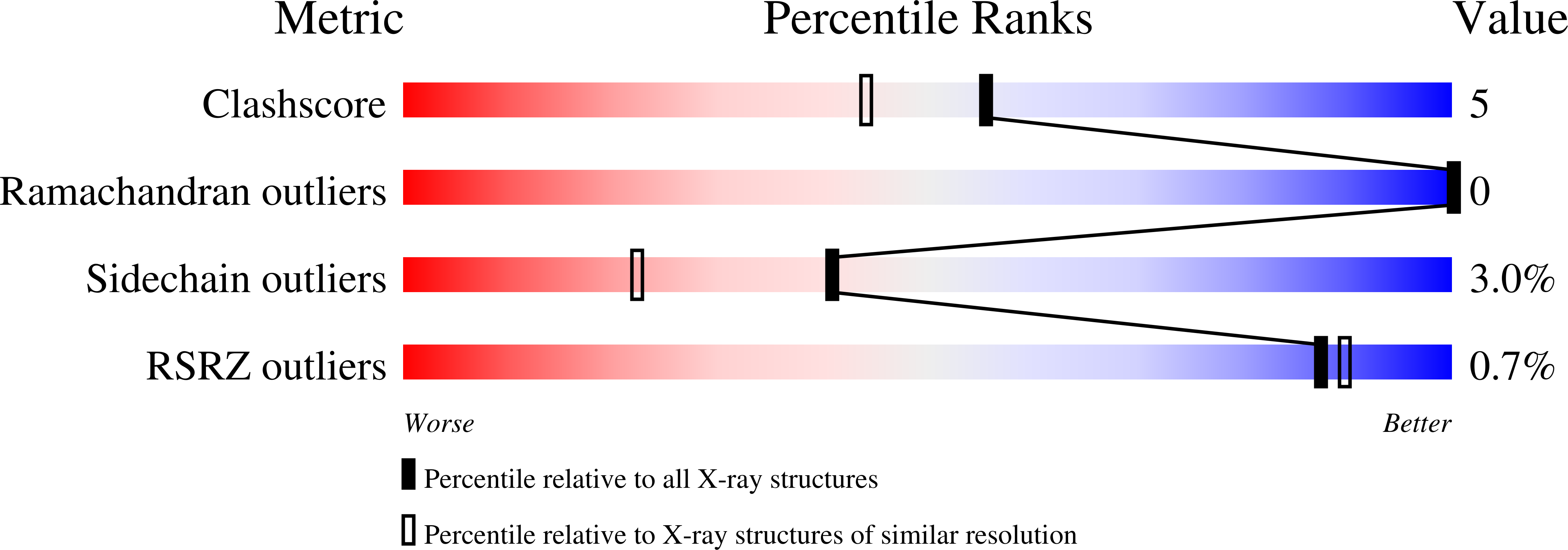
wwPDB Validation   3D Report Full Report


Ligand Structure Quality Assessment 


This is version 1.3 of the entry. See complete history


Literature

Crystal structure of oxygenated Scapharca dimeric hemoglobin at 1.7-A resolution.

Condon, P.J.Royer Jr., W.E.

(1994) J Biol Chem 269: 25259-25267

  • DOI: https://doi.org/10.2210/pdb1hbi/pdb
  • Primary Citation of Related Structures:  
    1HBI

  • PubMed Abstract: 

    The crystal structure of the cooperative dimeric hemoglobin from the blood clam Scapharca inaequivalvis has been determined in the oxygenated state and refined to an R-factor of 0.157 at 1.7-A resolution. The structure is very similar to the carbon monoxide-liganded form with subtle differences in ligand binding geometry. Oxygen binds to the heme iron in a bent conformation with Fe-O-O angles of 135 degrees and 150 degrees for the two subunits. These observed angles are lower than the equivalent angles in the carbon monoxide-liganded form and intermediate between the angles observed in structures of oxygenated sperm whale myoglobin and oxygenated human hemoglobin. This third high resolution structure of Scapharca dimeric hemoglobin permits a detailed analysis of the water structure in the highly hydrated interface between subunits.


  • Organizational Affiliation

    Program in Molecular Medicine, University of Massachusetts Medical Center, Worcester 01605.


Macromolecules
Find similar proteins by:  (by identity cutoff)  |  3D Structure
Entity ID: 1
MoleculeChains Sequence LengthOrganismDetailsImage
HEMOGLOBIN (OXY)
A, B
146Anadara inaequivalvisMutation(s): 0 
UniProt
Find proteins for P02213 (Anadara inaequivalvis)
Explore P02213 
Go to UniProtKB:  P02213
Entity Groups  
Sequence Clusters30% Identity50% Identity70% Identity90% Identity95% Identity100% Identity
UniProt GroupP02213
Sequence Annotations
Expand
  • Reference Sequence
Experimental Data & Validation

Experimental Data

  • Method: X-RAY DIFFRACTION
  • Resolution: 1.70 Å
  • R-Value Observed: 0.157 
  • Space Group: C 1 2 1
Unit Cell:
Length ( Å )Angle ( ˚ )
a = 93.2α = 90
b = 44β = 122
c = 83.5γ = 90
Software Package:
Software NamePurpose
X-PLORmodel building
PROLSQrefinement
X-PLORrefinement
X-PLORphasing

Structure Validation

View Full Validation Report


Ligand Structure Quality Assessment 


Entry History 

Deposition Data

Revision History  (Full details and data files)

  • Version 1.0: 1994-10-15
    Type: Initial release
  • Version 1.1: 2008-03-03
    Changes: Version format compliance
  • Version 1.2: 2011-07-13
    Changes: Version format compliance
  • Version 1.3: 2024-02-07
    Changes: Data collection, Database references, Derived calculations, Other