1GWS

hexadecaheme high molecular weight cytochrome Hmc from Desulfovibrio vulgaris Hildenborough


Experimental Data Snapshot

  • Method: X-RAY DIFFRACTION
  • Resolution: 2.40 Å
  • R-Value Free: 0.276 
  • R-Value Work: 0.201 

wwPDB Validation   3D Report Full Report


Ligand Structure Quality Assessment 


This is version 2.0 of the entry. See complete history


Literature

The Crystal Structure of the Hexadaca-Heme Cytochrome Hmc and a Structural Model of its Complex with Cytochrome C3

Czjzek, M.Elantak, L.Zamboni, V.Morelli, X.Dolla, A.Guerlesquin, F.Bruschi, M.

(2002) Structure 10: 1677

  • DOI: https://doi.org/10.1016/s0969-2126(02)00909-7
  • Primary Citation of Related Structures:  
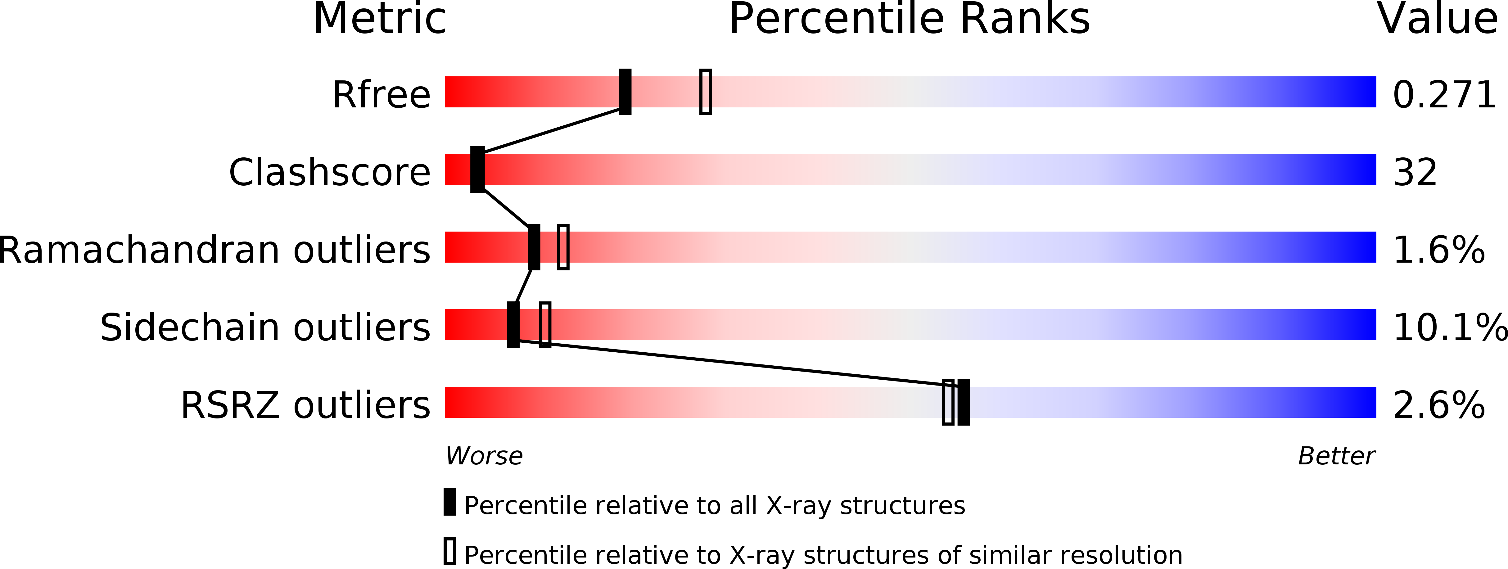
    1GWS

  • PubMed Abstract: 

    Sulfate-reducing bacteria contain a variety of multi-heme c-type cytochromes. The cytochrome of highest molecular weight (Hmc) contains 16 heme groups and is part of a transmembrane complex involved in the sulfate respiration pathway. We present the 2.42 A resolution crystal structure of the Desulfovibrio vulgaris Hildenborough cytochrome Hmc and a structural model of the complex with its physiological electron transfer partner, cytochrome c(3), obtained by NMR restrained soft-docking calculations. The Hmc is composed of three domains, which exist independently in different sulfate-reducing species, namely cytochrome c(3), cytochrome c(7), and Hcc. The complex involves the last heme at the C-terminal region of the V-shaped Hmc and heme 4 of cytochrome c(3), and represents an example for specific cytochrome-cytochrome interaction.


  • Organizational Affiliation

    Architecture et Fonction des Macromolécules Biologiques, IBSM-CNRS et Université Aix-Marseille I et II, 31 Chemin Joseph-Aiguier, 13402 Marseille cedex 20, France. czjzek@afmb.cnrs-mrs.fr


Macromolecules
Find similar proteins by:  (by identity cutoff)  |  3D Structure
Entity ID: 1
MoleculeChains Sequence LengthOrganismDetailsImage
HIGH-MOLECULAR-WEIGHT CYTOCHROME C545Nitratidesulfovibrio vulgaris str. HildenboroughMutation(s): 0 
UniProt
Find proteins for P24092 (Desulfovibrio vulgaris (strain ATCC 29579 / DSM 644 / NCIMB 8303 / VKM B-1760 / Hildenborough))
Explore P24092 
Go to UniProtKB:  P24092
Entity Groups  
Sequence Clusters30% Identity50% Identity70% Identity90% Identity95% Identity100% Identity
UniProt GroupP24092
Sequence Annotations
Expand
  • Reference Sequence
Small Molecules
Ligands 1 Unique
IDChains Name / Formula / InChI Key2D Diagram3D Interactions
HEC
Query on HEC

Download Ideal Coordinates CCD File 
B [auth A]
C [auth A]
D [auth A]
E [auth A]
F [auth A]
B [auth A],
C [auth A],
D [auth A],
E [auth A],
F [auth A],
G [auth A],
H [auth A],
I [auth A],
J [auth A],
K [auth A],
L [auth A],
M [auth A],
N [auth A],
O [auth A],
P [auth A],
Q [auth A]
HEME C
C34 H34 Fe N4 O4
HXQIYSLZKNYNMH-LJNAALQVSA-N
Experimental Data & Validation

Experimental Data

  • Method: X-RAY DIFFRACTION
  • Resolution: 2.40 Å
  • R-Value Free: 0.276 
  • R-Value Work: 0.201 
  • Space Group: P 62
Unit Cell:
Length ( Å )Angle ( ˚ )
a = 108.386α = 90
b = 108.386β = 90
c = 102.809γ = 120
Software Package:
Software NamePurpose
REFMACrefinement
DENZOdata reduction
SCALAdata scaling
SHARPphasing

Structure Validation

View Full Validation Report


Ligand Structure Quality Assessment 


Entry History 

Deposition Data

Revision History  (Full details and data files)

  • Version 1.0: 2003-02-13
    Type: Initial release
  • Version 1.1: 2011-07-13
    Changes: Advisory, Version format compliance
  • Version 2.0: 2019-09-25
    Changes: Advisory, Atomic model, Data collection, Derived calculations, Experimental preparation, Non-polymer description, Other, Structure summary