1GDL

CRYSTAL STRUCTURE OF FERRIC COMPLEXES OF THE YELLOW LUPIN LEGHEMOGLOBIN WITH ISOQUINOLINE AT 1.8 ANGSTROMS RESOLUTION (RUSSIAN)


Experimental Data Snapshot

  • Method: X-RAY DIFFRACTION
  • Resolution: 1.80 Å
  • R-Value Observed: 0.175 

wwPDB Validation   3D Report Full Report


This is version 1.4 of the entry. See complete history


Literature

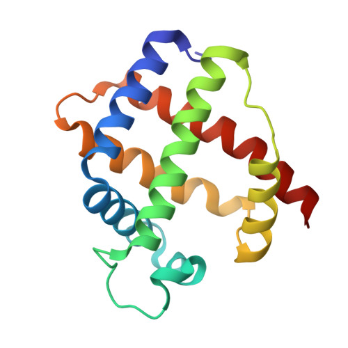
The binding of carbon monoxide and nitric oxide to leghaemoglobin in comparison with other haemoglobins.

Harutyunyan, E.H.Safonova, T.N.Kuranova, I.P.Popov, A.N.Teplyakov, A.V.Obmolova, G.V.Valnshtein, B.K.Dodson, G.G.Wilson, J.C.

(1996) J Mol Biol 264: 152-161

  • DOI: https://doi.org/10.1006/jmbi.1996.0630
  • Primary Citation of Related Structures:  
    1GDI, 1GDL

  • PubMed Abstract: 

    Haemoglobins have the ability to discriminate between oxygen and other diatomic molecules. To further understanding of this process the X-ray crystal structures of carbonmonoxy and nitrosyl-leghaemoglobin have been determined at 1.8 A resolution. The ligand geometry is discussed in detail and the controversial issue of bent versus linear carbon monoxide binding is addressed. The bond angle of 160 degrees for CO-leghaemoglobin is in conflict with recent spectroscopy results on myoglobin but is consistent with angles obtained for myoglobin X-ray crystal structures. In contrast to the numerous carbon monoxide studies, very little stereochemical information is available for the nitric oxide adduct of haemoglobin. This is provided by the X-ray structure of NO-leghaemoglobin, which conforms to expected geometry with an Fe-NO angle of 147 degrees and a lengthened iron-proximal histidine bond. Thus crystallographic evidence is given for the predicted weakening of this bond on the binding of nitric oxide.


  • Organizational Affiliation

    Institute of Crystallography, Russian Academy of Sciences, Moscow, Russia.


Macromolecules
Find similar proteins by:  (by identity cutoff)  |  3D Structure
Entity ID: 1
MoleculeChains Sequence LengthOrganismDetailsImage
LEGHEMOGLOBIN (NITROGEN MONOXY)153Lupinus luteusMutation(s): 0 
UniProt
Find proteins for P02240 (Lupinus luteus)
Explore P02240 
Go to UniProtKB:  P02240
Entity Groups  
Sequence Clusters30% Identity50% Identity70% Identity90% Identity95% Identity100% Identity
UniProt GroupP02240
Sequence Annotations
Expand
  • Reference Sequence
Small Molecules
Ligands 2 Unique
IDChains Name / Formula / InChI Key2D Diagram3D Interactions
HEM
Query on HEM

Download Ideal Coordinates CCD File 
B [auth A]PROTOPORPHYRIN IX CONTAINING FE
C34 H32 Fe N4 O4
KABFMIBPWCXCRK-RGGAHWMASA-L
NO
Query on NO

Download Ideal Coordinates CCD File 
C [auth A]NITRIC OXIDE
N O
ODUCDPQEXGNKDN-UHFFFAOYSA-N
Experimental Data & Validation

Experimental Data

  • Method: X-RAY DIFFRACTION
  • Resolution: 1.80 Å
  • R-Value Observed: 0.175 
  • Space Group: C 1 2 1
Unit Cell:
Length ( Å )Angle ( ˚ )
a = 93.47α = 90
b = 51.98β = 98.8
c = 38.32γ = 90
Software Package:
Software NamePurpose
PROLSQrefinement

Structure Validation

View Full Validation Report


Entry History 

Deposition Data

Revision History  (Full details and data files)

  • Version 1.0: 1995-02-27
    Type: Initial release
  • Version 1.1: 2008-03-24
    Changes: Version format compliance
  • Version 1.2: 2011-07-13
    Changes: Version format compliance
  • Version 1.3: 2017-11-29
    Changes: Derived calculations, Other
  • Version 1.4: 2024-02-07
    Changes: Data collection, Database references, Derived calculations