1FGG

CRYSTAL STRUCTURE OF 1,3-GLUCURONYLTRANSFERASE I (GLCAT-I) COMPLEXED WITH GAL-GAL-XYL, UDP, AND MN2+


Experimental Data Snapshot

  • Method: X-RAY DIFFRACTION
  • Resolution: 2.30 Å
  • R-Value Free: 0.215 
  • R-Value Work: 0.188 
  • R-Value Observed: 0.189 

wwPDB Validation   3D Report Full Report


This is version 2.1 of the entry. See complete history


Literature

Heparan/chondroitin sulfate biosynthesis. Structure and mechanism of human glucuronyltransferase I.

Pedersen, L.C.Tsuchida, K.Kitagawa, H.Sugahara, K.Darden, T.A.Negishi, M.

(2000) J Biol Chem 275: 34580-34585

  • DOI: https://doi.org/10.1074/jbc.M007399200
  • Primary Citation of Related Structures:  
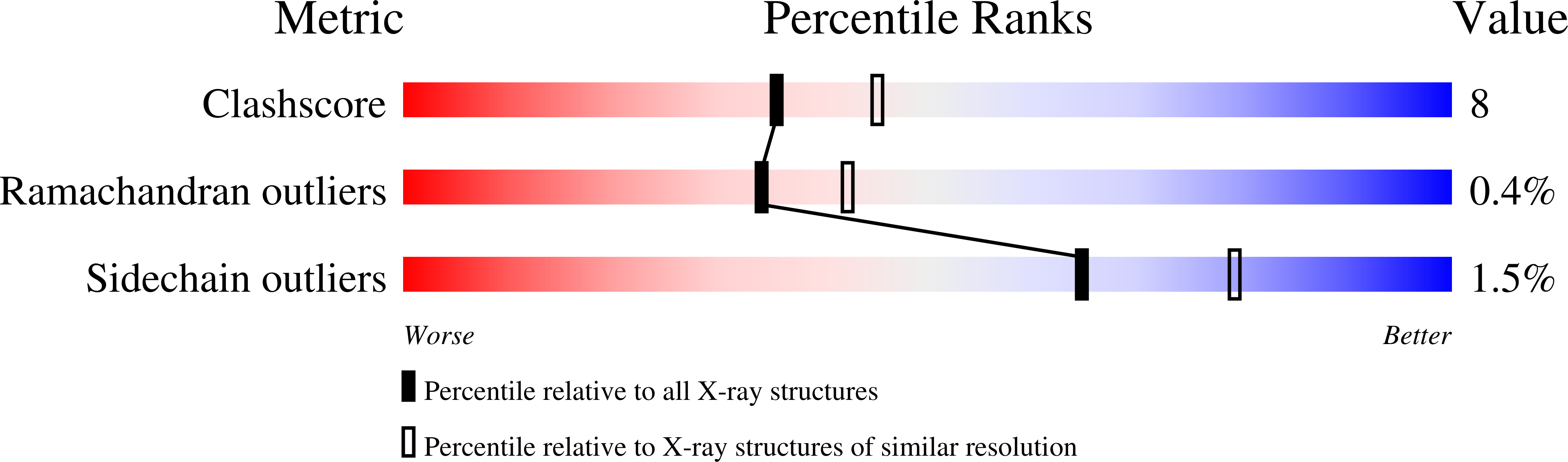
    1FGG

  • PubMed Abstract: 

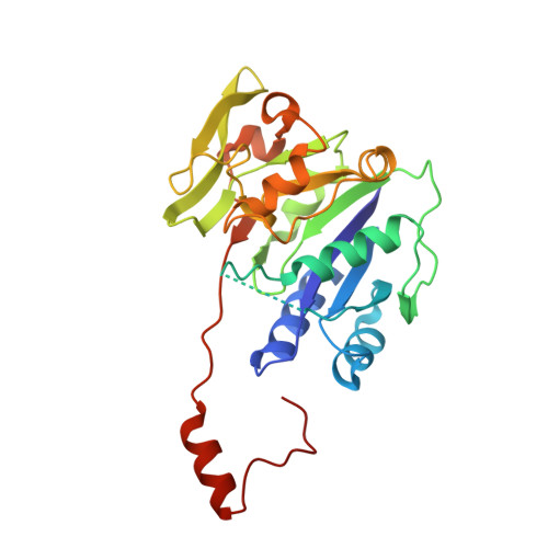
    Human beta1,3-glucuronyltransferase I (GlcAT-I) is a central enzyme in the initial steps of proteoglycan synthesis. GlcAT-I transfers a glucuronic acid moiety from the uridine diphosphate-glucuronic acid (UDP-GlcUA) to the common linkage region trisaccharide Gal beta 1-3Gal beta 1-4Xyl covalently bound to a Ser residue at the glycosaminylglycan attachment site of proteoglycans. We have now determined the crystal structure of GlcAT-1 at 2.3 A in the presence of the donor substrate product UDP, the catalytic Mn(2+) ion, and the acceptor substrate analog Gal beta 1-3Gal beta 1-4Xyl. The enzyme is a alpha/beta protein with two subdomains that constitute the donor and acceptor substrate binding site. The active site residues lie in a cleft extending across both subdomains in which the trisaccharide molecule is oriented perpendicular to the UDP. Residues Glu(227), Asp(252), and Glu(281) dictate the binding orientation of the terminal Gal-2 moiety. Residue Glu(281) is in position to function as a catalytic base by deprotonating the incoming 3-hydroxyl group of the acceptor. The conserved DXD motif (Asp(194), Asp(195), Asp(196)) has direct interaction with the ribose of the UDP molecule as well as with the Mn(2+) ion. The key residues involved in substrate binding and catalysis are conserved in the glucuronyltransferase family as well as other glycosyltransferases.


  • Organizational Affiliation

    Pharmacogenetic Section, Laboratory of Reproductive and Developmental Toxicology, NIEHS, National Institutes of Health, Research Triangle Park, North Carolina 27709, USA.


Macromolecules
Find similar proteins by:  (by identity cutoff)  |  3D Structure
Entity ID: 1
MoleculeChains Sequence LengthOrganismDetailsImage
GLUCURONYLTRANSFERASE I
A, B
261Homo sapiensMutation(s): 0 
UniProt & NIH Common Fund Data Resources
Find proteins for O94766 (Homo sapiens)
Explore O94766 
Go to UniProtKB:  O94766
PHAROS:  O94766
GTEx:  ENSG00000149541 
Entity Groups  
Sequence Clusters30% Identity50% Identity70% Identity90% Identity95% Identity100% Identity
UniProt GroupO94766
Sequence Annotations
Expand
  • Reference Sequence
Oligosaccharides

Help

Entity ID: 2
MoleculeChains Length2D Diagram Glycosylation3D Interactions
beta-D-galactopyranose-(1-3)-beta-D-galactopyranose
C
2N/A
Glycosylation Resources
GlyTouCan:  G95894VW
GlyCosmos:  G95894VW
GlyGen:  G95894VW
Experimental Data & Validation

Experimental Data

  • Method: X-RAY DIFFRACTION
  • Resolution: 2.30 Å
  • R-Value Free: 0.215 
  • R-Value Work: 0.188 
  • R-Value Observed: 0.189 
  • Space Group: P 1 21 1
Unit Cell:
Length ( Å )Angle ( ˚ )
a = 57.842α = 90
b = 48.219β = 92.79
c = 102.204γ = 90
Software Package:
Software NamePurpose
CNSrefinement
SCALEPACKdata scaling
CNSphasing

Structure Validation

View Full Validation Report


Entry History 

Deposition Data

Revision History  (Full details and data files)

  • Version 1.0: 2001-01-31
    Type: Initial release
  • Version 1.1: 2008-04-27
    Changes: Version format compliance
  • Version 1.2: 2011-07-13
    Changes: Version format compliance
  • Version 1.3: 2017-02-01
    Changes: Structure summary
  • Version 1.4: 2017-10-04
    Changes: Refinement description
  • Version 1.5: 2018-01-31
    Changes: Experimental preparation
  • Version 2.0: 2020-07-29
    Type: Remediation
    Reason: Carbohydrate remediation
    Changes: Atomic model, Data collection, Database references, Derived calculations, Structure summary
  • Version 2.1: 2024-03-13
    Changes: Data collection, Database references, Source and taxonomy, Structure summary