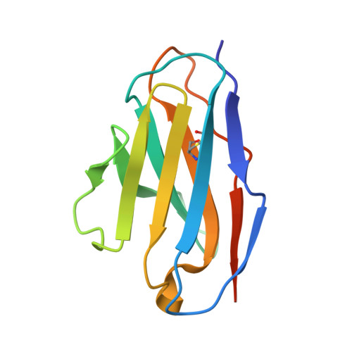
Antiferritin VL homodimer binds human spleen ferritin with high specificity
Nymalm, Y., Kravchuk, Z., Salminen, T., Chumanevich, A.A., Dubnovitsky, A.P., Kankare, J., Pentikainen, O., Lehtonen, J., Arosio, P., Martsev, S., Johnson, M.S.(2002) J Struct Biol 138: 171-186
- PubMed: 12217656
- DOI: https://doi.org/10.1016/s1047-8477(02)00015-1
- Primary Citation of Related Structures:
1F6L - PubMed Abstract:
The antiferritin variable light domain (VL) dimer binds human spleen ferritin ( approximately 85% L subunits) but with approximately 50-fold lower affinity, K(a)=4 x 10(7) x M(-1), than the parent F11 antibody (K(a)=2.1 x 10(9) x M(-1)). The VL dimer does not recognize either rL (100% L subunits) or rH (100% H subunits) human ferritin, whereas the parent antibody recognizes rL-ferritin. To help explain the differences in ferritin binding affinities and specificities, the crystal structure of the VL domain (2.8A resolution) was determined by molecular replacement and models of the antiferritin VL-VH dimer were made on the basis of antilysozyme antibody D1.3. The domain interface is smaller in the VL dimer but a larger number of interdomain hydrogen bonds may prevent rearrangement on antigen binding. The antigen binding surface of the VL dimer is flatter, lacking a negatively charged pocket found in the VL-VH models, contributed by the CDR3 loop of the VH domain. Loop CDR2 (VL dimer) is located away from the antigen binding site, while the corresponding loop of the VH domain would be located within the antigen binding site. Together these differences lead to 50-fold lower binding affinity in the VL dimer and to more restricted specificity than is seen for the parent antibody.
Organizational Affiliation:
Department of Biochemistry and Pharmacy, Abo Akademi University, P.O. Box 66, FIN-20521, Turku, Finland.