The 1.9 A crystal structure of the "as isolated" rubrerythrin from Desulfovibrio vulgaris: some surprising results.
Sieker, L.C., Holmes, M., Le Trong, I., Turley, S., Liu, M.Y., LeGall, J., Stenkamp, R.E.(2000) J Biol Inorg Chem 5: 505-513
- PubMed: 10968622
- DOI: https://doi.org/10.1007/pl00021450
- Primary Citation of Related Structures:
1DVB - PubMed Abstract:
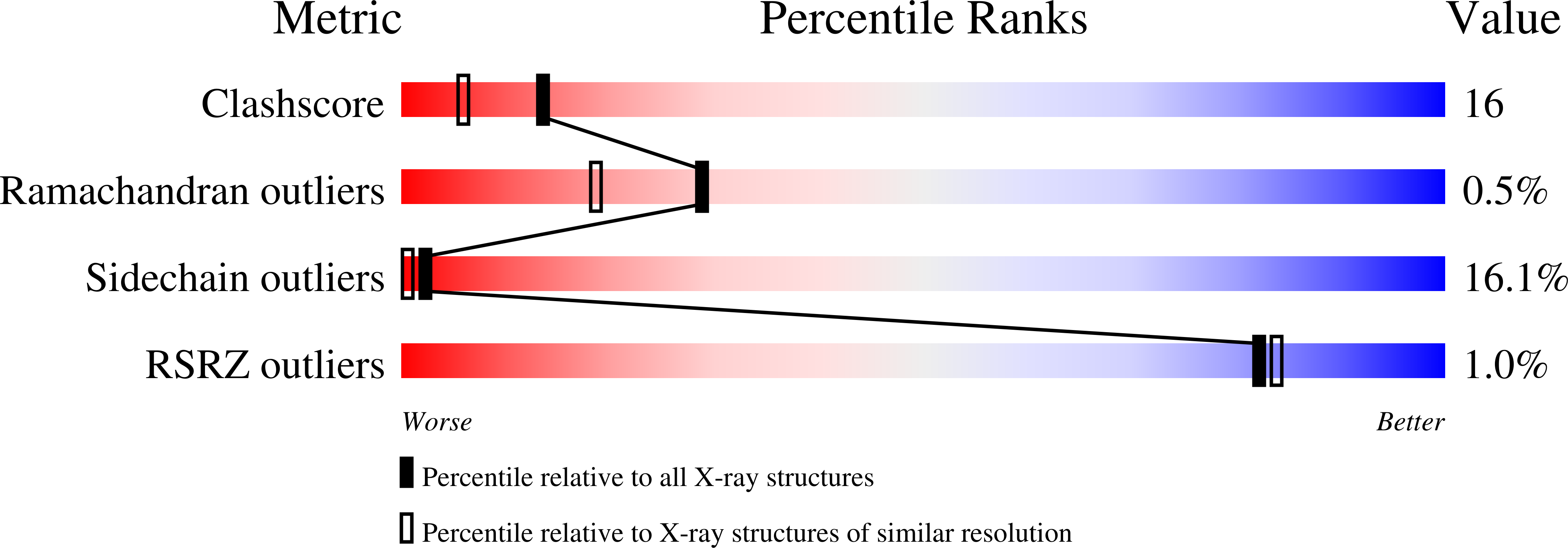
Rubrerythrin is a non-heme iron dimeric protein isolated from the sulfate-reducing bacterium Desulfovibrio vulgaris. Each monomer has one mononuclear iron center similar to rubredoxin and one dinuclear metal center similar to hemerythrin or ribonucleotide reductase. The 1.88 A X-ray structure of the "as isolated" molecule and a uranyl heavy atom derivative have been solved by molecular replacement techniques. The resulting model of the native "as isolated" molecule, including 164 water molecules, has been refined giving a final R factor of 0.197 (R(free) = 0.255). The structure has the same general protein fold, domain structure, and dimeric interactions as previously found for rubrerythrin [1, 2], but it also has some interesting undetected differences at the metal centers. The refined model of the protein structure has a cis peptide between residues 78 and 79. The Fe-Cys4 center has a previously undetected strong seventh N-H...S hydrogen bond in addition to the six N-H...S bonds usually found in rubredoxin. The dinuclear metal center has a hexacoordinate Fe atom and a tetracoordinate Zn atom. Each metal is coordinated by a GluXXHis polypeptide chain segment. The Zn atom binds at a site distinctly different from that found in the structure of a diiron rubrerythrin. Difference electron density for the uranyl derivative shows an extremely large peak adjacent to and replacing the Zn atom, indicating that this particular site is capable of binding other atoms. This feature/ability may give rise to some of the confusing activities ascribed to this molecule.
Organizational Affiliation:
Department of Biological Structure and Biomolecular Structure Center, University of Washington, Seattle 98195-7420, USA.


