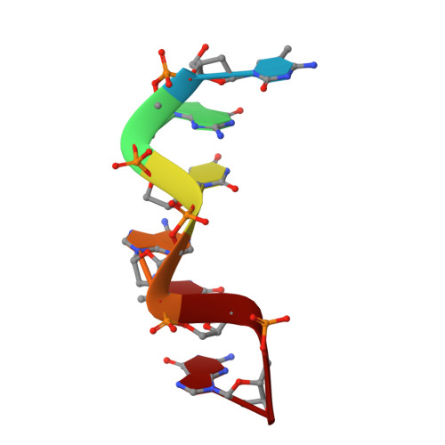
Base-specific binding of copper(II) to Z-DNA. The 1.3-A single crystal structure of d(m5CGUAm5CG) in the presence of CuCl2.
Geierstanger, B.H., Kagawa, T.F., Chen, S.L., Quigley, G.J., Ho, P.S.(1991) J Biol Chem 266: 20185-20191
- PubMed: 1939079
- DOI: https://doi.org/10.2210/pdb1d40/pdb
- Primary Citation of Related Structures:
1D40 - PubMed Abstract:
The single crystal structure of d(m5CGUAm5CG) soaked with copper(II) chloride was solved to atomic (1.3 A) resolution to study the base specificity of copper binding to double-stranded DNA. In the present copper(II) chloride-soaked structure, four crystallographically unique copper(II) complexes were observed bound to five of the six purine bases in the hexamer duplex. Covalent copper(II) binding occurred at N-7 of all four guanine bases and at one of the two adenine bases in the DNA duplex. Copper binding was not observed at the position (Ade4) located in an open solvent channel, whereas the second adenine site (Ade10) shared a complex with a guanine residue (Gua12) of a neighboring symmetry-related hexamer. The coordination geometries and distribution of these copper(II) complexes at the guanine bases in the crystal were comparable to the analogous sites in the isomorphous copper(II) chloride-soaked d(CGCGCG) crystal (Kagawa, T., Geierstanger, B. H., Wang, A. H.-J., and Ho, P.S. (1991) J. Biol. Chem. 266, 20175-20184). Thus, the decreased copper(II) binding affinity for Ade4 was not an artifact of crystal packing, but is intrinsic to the chemical properties of this purine base in duplex DNA. This suggests that the adenine bases in dilute solutions of Z-DNA and more generally other duplex DNA conformations are not susceptible to copper(II) modification. Thus, preferential copper(II) binding at guanine bases over adenine bases in double-stranded DNA may explain the observed specificity of copper(II)-induced oxidative DNA damage near guanine residues (Yamamoto, K., and Kawanishi, S. (1989) J. Biol. Chem. 264, 15435-15440; Sagripanti, J.-L., and Kraemer, K. H. (1989) J. Biol. Chem. 264, 1729-1734). The sharing of a single copper(II) complex by Ade10 and Gua12 of an adjacent hexamer suggests that additional and perhaps specific DNA-DNA interactions, as may be found in the densely packed environment of the nuclear matrix in the cell, may render N-7 of adenine bases prone to copper(II) modification.
Organizational Affiliation:
Department of Biochemistry and Biophysics, Oregon State University, Corvallis 97331-6503.